
Effect of hydroxyapatite bioceramics on the growth of osteoblasts and HIF-α/VEGF signal axis in partial hypoxia environment in vitro
Abstract
BACKGROUND:
Hydroxyapatite bioceramic is a kind of bone implant commonly used in oral clinic treatment. In the early stage of tissue repair, cells will suffer hypoxic due to the interruption of blood supply.
OBJECTIVE:
Studying the expression of osteoblasts in hypoxic environment will help us to understand the expression and response mechanism of osteoblasts at the implantation site of hydroxyapatite in the early stage of hypoxia.
METHODS:
MG63 osteoblast cell line was used in this study. The cells of normal group were incubated under normal oxygen and hydroxyapatite ceramics condition. The cells of hypoxia group were incubated under hypoxia (37
RESULTS:
Compared to the normal group, the cells of hypoxia group showed a dramatically higher proliferation ability, especially at 48 h (
CONCLUSIONS:
Under hypoxia condition, hydroxyapatite bioceramics can promote the proliferation of MG63 osteoblasts, elevate the activity of alkaline phosphatase and upregulate HIF-
1.Introduction
Hydroxyapatite bioceramics are rich in minerals, one of the most important components is hydroxy-apatite crystal [Ca
In the process of hydroxyapatite bioceramic filling treatment, the oral alveolar bone environment is not a complete oxygen environment. In the early stage of bone defects, there is not enough oxygen for cell growth due to the limitation of local microcirculation. At this time, the adaptation of cells to hypoxia and the promotion of vascular intercellular response are crucial for bone regeneration and wound healing [4]. HIF-
Human osteosarcoma cells (MG63) are osteoblast like cells and belong to osteoblast cell line. Due to the limitation of passage times and cell source of primary osteoblasts, we selected osteoblast like cell line to replace primary cells in our study. In this study, we found that, under hypoxia condition, hydroxyapatite bioceramics can promote the proliferation of MG63 osteoblasts, elevate the activity of alkaline phosphatase and upregulate HIF-
2.Materials and method
2.1Cell culture
Human osteosarcoma cells (MG63) were cultured in Dulbecco’s Modified Eagle Medium (DMEM) (Gibco, AE29040273) plus 10% fetal bovine serum (FBS) (Gibco, 2045677RP) and 1% penicllin-streptomcyin (P/S) (Gibco, 2068817). Under normal conditions, all cells were cultured at 37
2.2Preparation of hydroxyapatite bioceramics
Hydroxyapatite bioceramics were synthetized by Beijing Yihuajian science and Trade Co., Ltd., China, and extracted according to the scheme in part 12 of ISO10993.12-2008 biological evaluation of medical devices. The stocking concentration of hydroxyapatite bioceramic is 0.2 g/ml. Briefly, to dissolve the hydroxyapatite bioceramics, 0.2 g hydroxyapatite bioceramics were added into 1 ml intact medium and extracted in inert container at 37
2.3Cell proliferation assay
MG63 cells were cultured in serum-free medium in 96 well plate for 24 hours. The number of cells per well was 1
2.4Apoptosis assay
MG63 cells were plated at 6 cm dish with the density of 4
2.5ALP activity assay
MG63 cells were plated at 6 cm dish with the density of 4
2.6Western blot analysis
The cell lysates of MG63 cells were lyzed in ice-cold RIPA buffer. Protein concentrations of the cell lysates were determined by Bradford Protein assay (BCA). 20
2.7Statistical analysis
All the represent data from three independent experiments. The results were presented as the mean
3.Results
3.1Hydroxyapatite bioceramics promoted cell proliferation under hypoxia condition
The cell proliferation activity was evaluated by CCK8 assay in the co-culture system at 4 h, 8 h, 24 h and 48 h, respectively. The results showed that under hypoxia condition, hydroxyapatite bioceramics can promote the proliferation of MG63 osteoblasts (
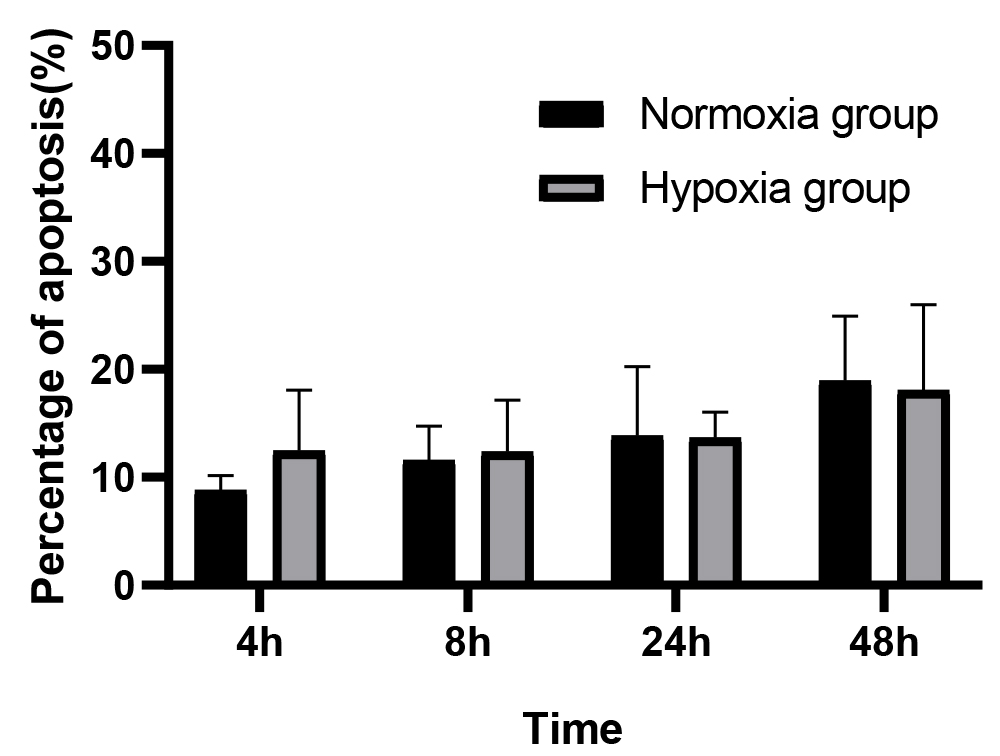
3.2Hydroxyapatite bioceramics showed no obvious effect on apoptosis
The apoptosis assay was conducted by Annexin V-FITC and PI double staining, the result of which was read out by flow cytometer. The result showed that there was no significant difference in apoptosis between normal group and hypoxia group at 4 h, 8 h, 24 h and 48 h, respectively (Fig. 2). This may be due to the adaptability of cells in hydroxyapatite co-culture system.
Table 1
ALP activity protein in each group (per mg)
| Group | Mean | |
|---|---|---|
| Normal group 24 h | 2.823 | |
| Hypoxia group 24 h* | 3.555 | 0.010 |
| Normal group 48 h | 2.667 | |
| Hypoxia group 48 h* | 3.188 | 0.020 |
Figure 1.
Hydroxyapatite bioceramics promoted cell proliferation under hypoxia condition.
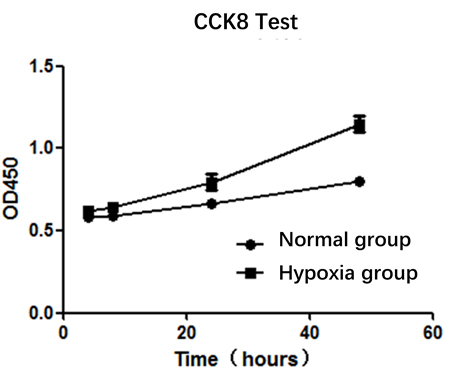
Figure 2.
Hydroxyapatite bioceramics showed no obvious effect on apoptosis.
3.3Hydroxyapatite bioceramics elevated ALP activity under hypoxia condition
Alkaline phosphatase (ALP) is a specific marker of osteoblast maturation, and is a common index to evaluate the secretion function of osteoblasts. In this study, the alkaline phosphate activity of MG63 osteoblast like cells was detected at 24 h and 48 h. The data from Table 1 showed that the activity of ALP in hypoxia group was higher than that in normal group (
Figure 3.
Hydroxyapatite bioceramics upregulated the protein levle of HIF-VEGF signal axis.
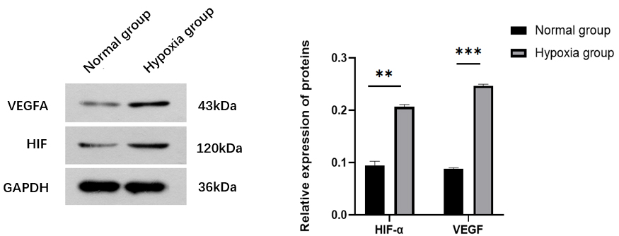
3.4Hydroxyapatite bioceramics upregulated the protein levle of HIF-VEGF signal axis
Mechanically, as shown in Fig. 3, the western blot result showed that after treatment with hydroxyapatite bioceramics for 24 h, the protein level of both HIF-
4.Conclusion
Hydroxyapatite bioceramics is a kind of bone implant material, and is commonly used in oral clinic. Hydroxyapatite bioceramics account for 60% of the extracellular matrix of the skeleton, and its chemical composition and crystal structure are very similar to the skeleton [7]. It not only supports the attachment of osteoblasts to bone, but also has strong osteoinductive properties. Studies have proved that it shows good osteogenic ability in the aspects of maxillary sinus wall defect, tissue defect after tumor removal or bone absorption around implant [8, 9]. In tissue engineering, cells can sense the changes of external conditions through their own biological signals and adapt to the changes of external materials and environment. A large number of in vitro experiments show that hydroxyapatite has good differentiation and proliferation effects on osteoblasts and stromal cells [10, 11, 12]. In the preparation of some tissue engineering scaffolds, hydroxyapatite is often used to promote the growth of bone cells [13, 14]. In the early stage of tissue repair, tissue cells will suffer partial hypoxic microenvironment in bone defect site due to blood supply interruption [15]. Studying the expression of osteoblasts in partial hypoxia environment will help us understand the behavior and mechanism of osteoblasts induced by partial hypoxia in the presence of hydroxyapatite bioceramics, so as to help us understand the cellular reaction and improve the properties of materials.
In this study we confirmed that under hypoxia condition, hydroxyapatite bioceramics can promote the proliferation of MG63 osteoblasts. Consistently, the cell proliferation rate increased much more significantly along with the extension of cell culture time (Fig. 1). However, it showed no effects on apoptosis, which may be related to the adaptability of cells in hydroxyapatite co culture system. ALP is a biochemical and histological marker to identify osteoblasts, and its activity reflects the differentiation and functional status of osteoblasts to a certain extent [16]. We found that the ALP activity of osteoblasts in hypoxia group was higher than that in normoxia group at 24 h and 48 h, indicating that the differentiation ability of osteoblasts and the bone metabolism of osteoblast like cells under hypoxia condition are more active than that in normoxia condition. Hypoxia inducible factor (HIF) is a heterodimeric protein composed of the constitutive subunit HIF-1
We found that hydroxyapatite ceramics can promote cell proliferation and differentiation in the early stage of culture under hypoxia condition (37
Conflict of interest
The authors state that there is no conflict of interest related to this article.
References
[1] | Azizi F, Heidari F, Fahimipour F, Sajjadnejad M, Vashaee D, Tayebi L. Evaluation of mechanical and biocompatibility properties of hydroxyapatite/manganese dioxide nanocomposite scaffolds for bone tissue engineering application. International Journal of Applied Ceramic Technology. (2020) ; 17: (5). |
[2] | Gradinaru S, Popescu V, Leasu C, Pricopie S, Yasin S, Ciuluvica R, Ungureanu E. Hydroxyapatite ocular implant and non-integrated implants in eviscerated patients. J Med Life. (2015) Jan-Mar; 8: (1): 90-3. |
[3] | Sakamoto Y, Sakamoto T, Ishii T, Kishi K. Assessment of bioabsorbable hydroxyapatite for secondary bone grafting in unilateral alveolar clefts. Cleft Palate Craniofac J. (2020) Jan; 57: (1): 114-117. |
[4] | Yellowley CE, Genetos DC. Hypoxia signaling in the skeleton: Implications for bone health. Curr Osteoporos Rep. (2019) Feb; 17: (1): 26-35. |
[5] | Branco-Price C, Evans CE, Johnson RS. Endothelial hypoxic metabolism in carcinogenesis and dissemination: HIF-A isoforms are a NO metastatic phenomenon. Oncotarget. (2013) Dec; 4: (12): 2567-76. |
[6] | Hu K, Olsen BR. Osteoblast-derived VEGF regulates osteoblast differentiation and bone formation during bone repair. J Clin Invest. (2016) Feb; 126: (2): 509-26. |
[7] | Guillen-Romero LD, Oropeza-Guzmán MT, López-Maldonado EA, Iglesias AL, Paz-González JA, Ng T, Serena-Gómez E, Villarreal-Gómez LJ. Synthetic hydroxyapatite and its use in bioactive coatings. J Appl Biomater Funct Mater. (2019) Jan-Mar; 17: (1). |
[8] | Khaled H, Atef M, Hakam M. Maxillary sinus floor elevation using hydroxyapatite nano particles vs tenting technique with simultaneous implant placement: A randomized clinical trial. Clin Implant Dent Relat Res. (2019) Dec; 21: (6): 1241-1252. |
[9] | Zhang K, Zhou Y, Xiao C, Zhao W, Wu H, Tang J, Li Z, Yu S, Li X, Min L, Yu Z, Wang G, Wang L, Zhang K, Yang X, Zhu X, Tu C, Zhang X. Application of hydroxyapatite nanoparticles in tumor-associated bone segmental defect. Sci Adv. (2019) Aug 2; 5: (8). |
[10] | Ersoy B, Bayramiçli M, Ercan F, Şrinoıglu H, Turan P, Numanoğlu A. Comparison of bone prefabrication with vascularized periosteal flaps, hydroxyapatite, and bioactive glass in rats. J Reconstr Microsurg. (2015) May; 31: (4): 291-9. |
[11] | Canettieri AC, Colombo CE, Chin CM, Faig-Leite H. Femur bone repair in ovariectomized rats under the local action of alendronate, hydroxyapatite and the association of alendronate and hydroxyapatite. Int J Exp Pathol. (2009) Oct; 90: (5): 520-6. |
[12] | Vamze J, Pilmane M, Skagers A. Biocompatibility of pure and mixed hydroxyapatite and α-tricalcium phosphate implanted in rabbit bone. J Mater Sci Mater Med. (2015) Feb; 26: (2): 73. |
[13] | Ramesh N, Moratti SC, Dias GJ. Hydroxyapatite-polymer biocomposites for bone regeneration: A review of current trends. J Biomed Mater Res B Appl Biomater. (2018) Jul; 106: (5): 2046-2057. |
[14] | Chen P, Liu L, Pan J, Mei J, Li C, Zheng Y. Biomimetic composite scaffold of hydroxyapatite/gelatin-chitosan core-shell nanofibers for bone tissue engineering. Mater Sci Eng C Mater Biol Appl. (2019) Apr; 97: : 325-335. |
[15] | Zuo X, Li J, Han X, Liu X, He H. Hypoxia inducible factor-1 α. Prevention and Treatment of Oral Diseases, (2021) ; 29: (7): 449-455. |
[16] | Topal İ, Gümüş B. Can Bone-Specific Alkaline Phosphatase and Osteocalcine Levels Be Used to Determine the Age in Children? Am J Forensic Med Pathol. (2020) Sep; 41: (3): 182-187. |
[17] | Branco-Price C, Evans CE, Johnson RS. Endothelial hypoxic metabolism in carcinogenesis and dissemination: HIF-A isoforms are a NO metastatic phenomenon. Oncotarget. (2013) Dec; 4: (12): 2567-76. |
[18] | Zhang S, Yuan Y, Zou J. Hypoxia inducible factor 1 α Progress in the regulation of bone metabolism and angiogenesis in bone microenvironment. Chinese Journal of Osteoporosis. (2020) ; 26: (8): 1201-1206. |
[19] | Debnath S, Mukherjee A, Saha D, Dash J, Chatterjee TK. Poly-l-Lysine inhibits VEGF and c-Myc mediated tumor-angiogenesis and induces apoptosis in 2D and 3D tumor microenvironment of both MDA-MB-231 and B16F10 induced mice model. International Journal of Biological Macromolecules. (2021) ; 183. |


