
Preliminary study on the electromagnetic field treatment of osteoporosis in rats
Abstract
OBJECTIVE:
In our study, the influence of PEMF on skeleton morphology and bone metabolism on rats with disuse osteoporosis was investigated, and the possibility of using it for the treatment of disuse osteoporosis was explored.
METHODS:
The rats in the ALN group were treated with alendronate, and the rats in the PEMF group were exposed to pulsed electromagnetic fields (3.82 mT, 10 Hz) for 40 mind
RESULTS:
The bone structural mechanical indices and material mechanical indices of the right femur in all groups of mice during weeks 2 and 4 were decreased. At week 8 the bone structural mechanical index and maximum stress of the right femur in the ALN group were markedly raised compared with the CON group (
CONCLUSION:
From the experimental time interval analysis, PEMF can improve the mechanical stability of bone structure more gently and permanently than alendronate.
1.Introduction
Disuse osteoporosis is common in patients who have been in bed for a long time as well as in astronauts who perform space missions, and is marked by the obliteration of trabecular bony structure and thinning of the cortical bone [1]. Reduction of mechanical stress, augmentation of osteoclast-facilitated bone resorption as well as inhibition of osteoblast-mediated bone generation are important contributors towards disuse osteoporosis [2]. Given our aging population and increasing dependence on mechanical tools, disuse osteoporosis has been a clinical entity of growing importance. Numerous experimental studies have directly or indirectly demonstrated that pulsed electromagnetic field therapy (PEMF) affects the potential of the Haval tube system, improving bone formation and remodeling [3]. However, there is no consensus regarding the causes of bone formation and remodeling in the literature. Thus, to clarify these contradictory findings, we established a rat tibia-tail fixation model to study bone morphology, bone biomechanics, and changes in the concentrations of bone morphogenetic protein-2 (BMP-2) and bone Gla-protein (BGP, osteocalcin) to investigate the efficacy of PEMF on disuse osteoporosis as a possible treatment method.
2.Materials and methods
2.1Animals
Our study was performed on SPF grade female rats (Sprague Dawley) that weighed between 250–280 g and were four weeks of age, and were supplied by the Jilin University (Certificate no. SCXK-Ji 2019-0008). All experimental procedures were reviewed by the Animal Ethics Committee of the First Hospital of Harbin.
2.2The PEMF system
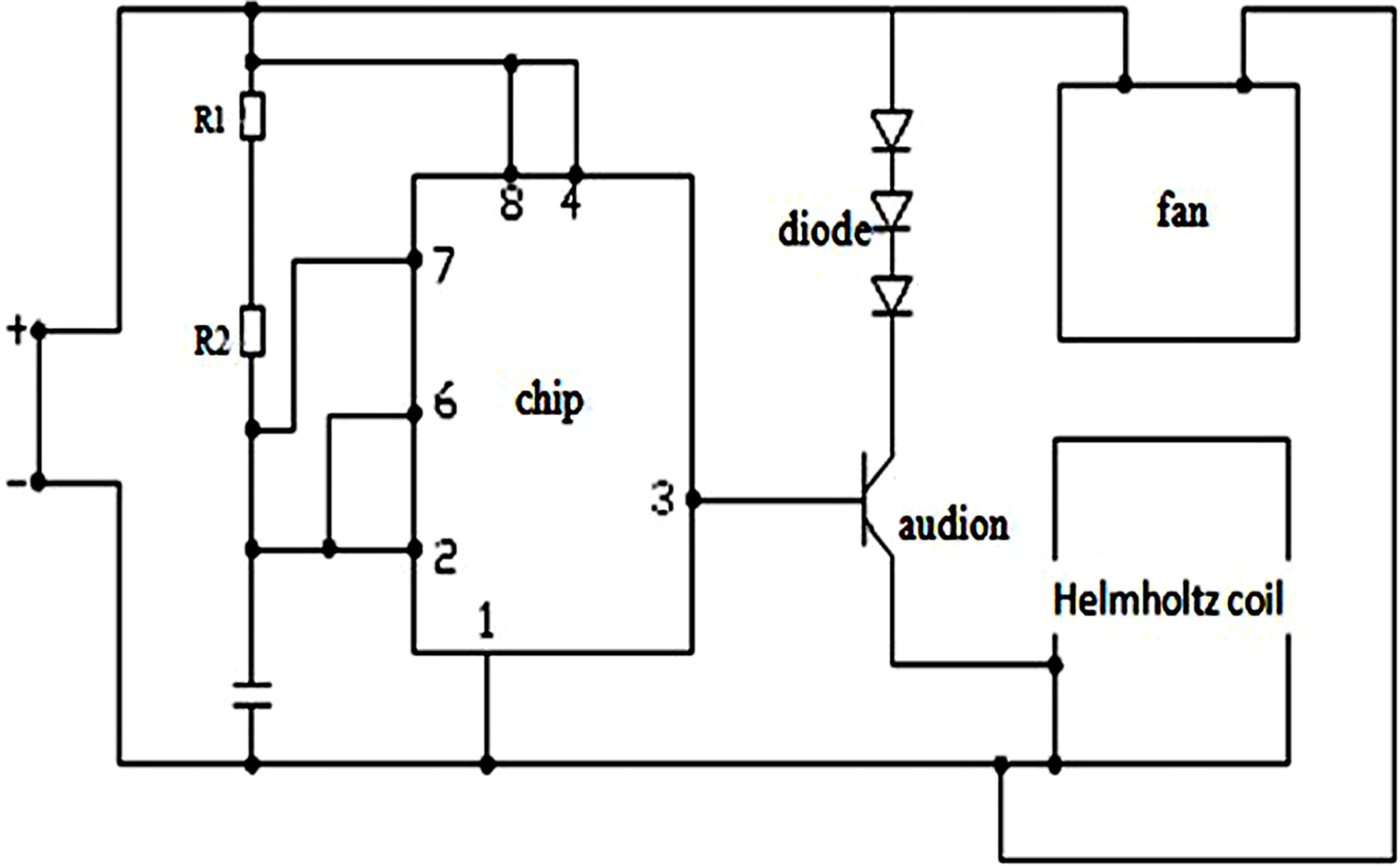
The current study utilizes Helmholtz coils and waveform generators in order to develop a PEMF exposure system. 240 mm diameter Helmholtz coils with 200 turns of copper wire diameter 0.9 mm were utilized to administer PEMF with 10-Hz repetitive single pulse waveforms produced by a waveform generator (Fig. 1).
Figure 1.
PEMF circuit schematic diagram.
2.3PEMF administration protocol and preparation of mice models
Four groups of rats were created based on random sampling of body mass (
PEMF irradiation treatment was administered to rats with parameters of 10 Hz and 3.82 mT in PEMF group for 40 min while those in the ALN group were administered alendronate sodium at a dose of 1 mg
2.4Bone tissue biomechanics, morphometry, and detection of serum BMP-2 and GDP concentrations
Five rats were sacrificed at 2, 4, 8, and 12 weeks of treatment for the detection of bone biomechanics, morphometry, and serum BMP-2 and BGP concentrations. Right ventricular blood samples were harvested, subjected to 3000 rpm centrifugation, and kept at
The three-point bending test was performed (ZWICK electronic universal testing machine model Z010, Harbin Institute of Technology Mechanics Experimental Center, China), with a loading speed of 2 mm
Four % paraformaldehyde was used for right tibia fixation prior to sectioning one-fifth of the sample. Bone morphology was assessed by electron microscopy (AMRAY 1000B scanning electron microscope, USA). ALN could markedly increase the total bone surface area (T-Ar), trabecular bone area (Tb-Ar) and trabecular circumference (Th-Pm). Subsequently, calculate the parameters according to the formula: Trabecular bone average interval width (Tb-Sp)
The rest of the tibia samples were subjected to immunohistochemistry (Beijing Bioss, China) in order to quantify BMP-2 expression.
2.5Statistical analysis
Data analysis was performed using SPSS statistics 25.0 software. One-way ANOVA was used to compare multiple sets of data, with a
3.Results
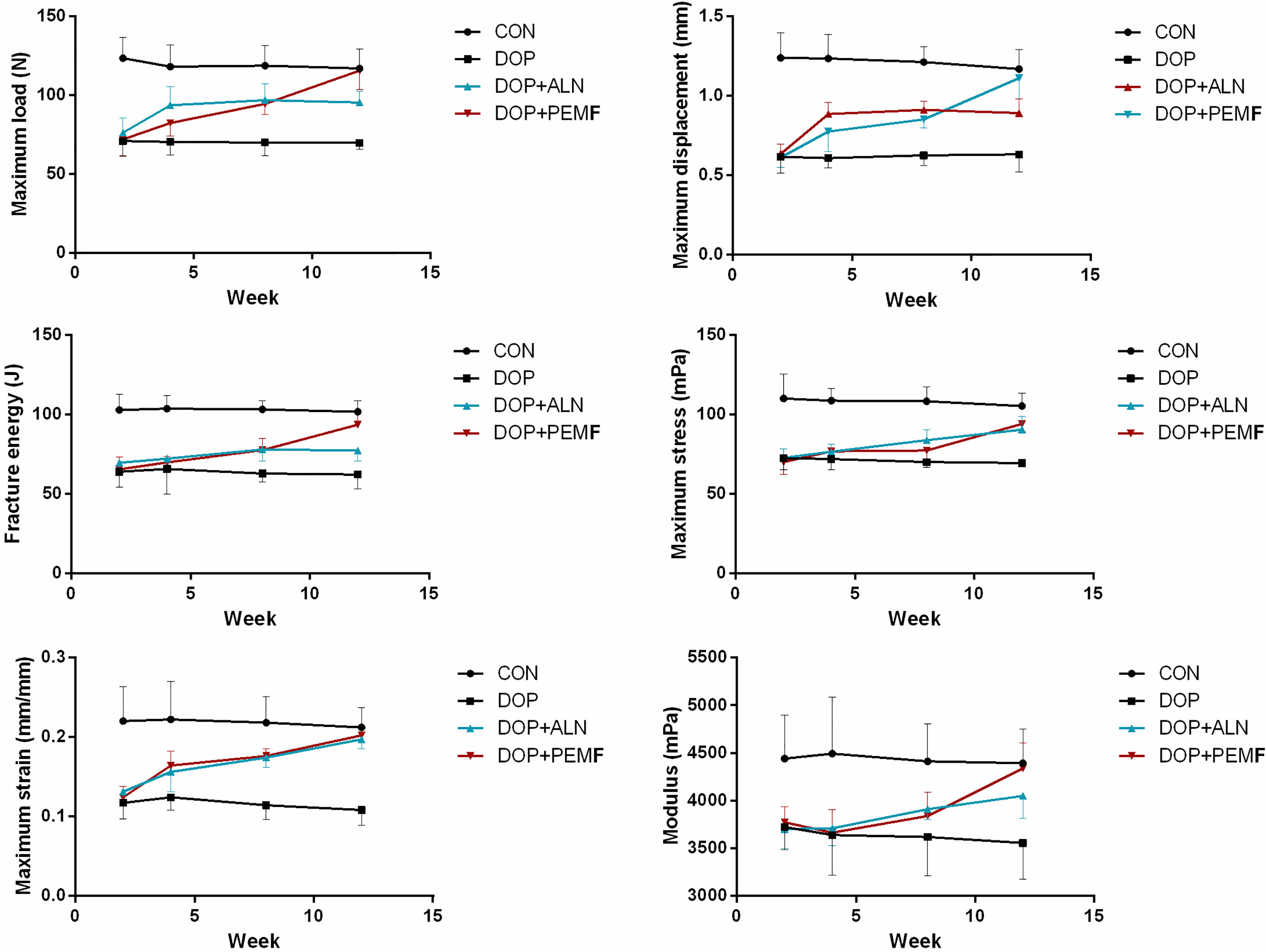
3.1PEMF improves the structural mechanics and mechanical parameters of femur in DOP rats
The femoral structural mechanics and material mechanics of rats across all groups were decreased (
At week 12, in comparison with the model group, the structural mechanical indices in the ALN group were significantly raised (
Figure 2.
Comparison of the biomechanical parameters in each group.
3.2PEMF increased femoral Tb-N and %Tb-Ar and decreased Tb-Sp in DOP rats
Both the %Tb-Ar and Tb-N in ALN group were markedly raised in contrast to the DOP group following two and four weeks of treatment, whereas the Tb-Sp was suppressed (
Likewise, at four weeks, the Tb-N and %Tb-Ar of the ALN group were markedly raised when compared to the DOP group, whereas Tb-Sp was obviously reduced (
At 8 weeks, the index parameters Tb-N and %Tb-Ar in both the ALN and PEMF groups were markedly raised while the Tb-Sp was suppressed significantly when compared to the DOP group (
At 12 weeks, the Tb-N and %Tb-Ar in both the ALN and the PEMF groups were significantly increased (
Figure 3.
Variation in the bone histomorphometry following 4 weeks of PEMF treatment.
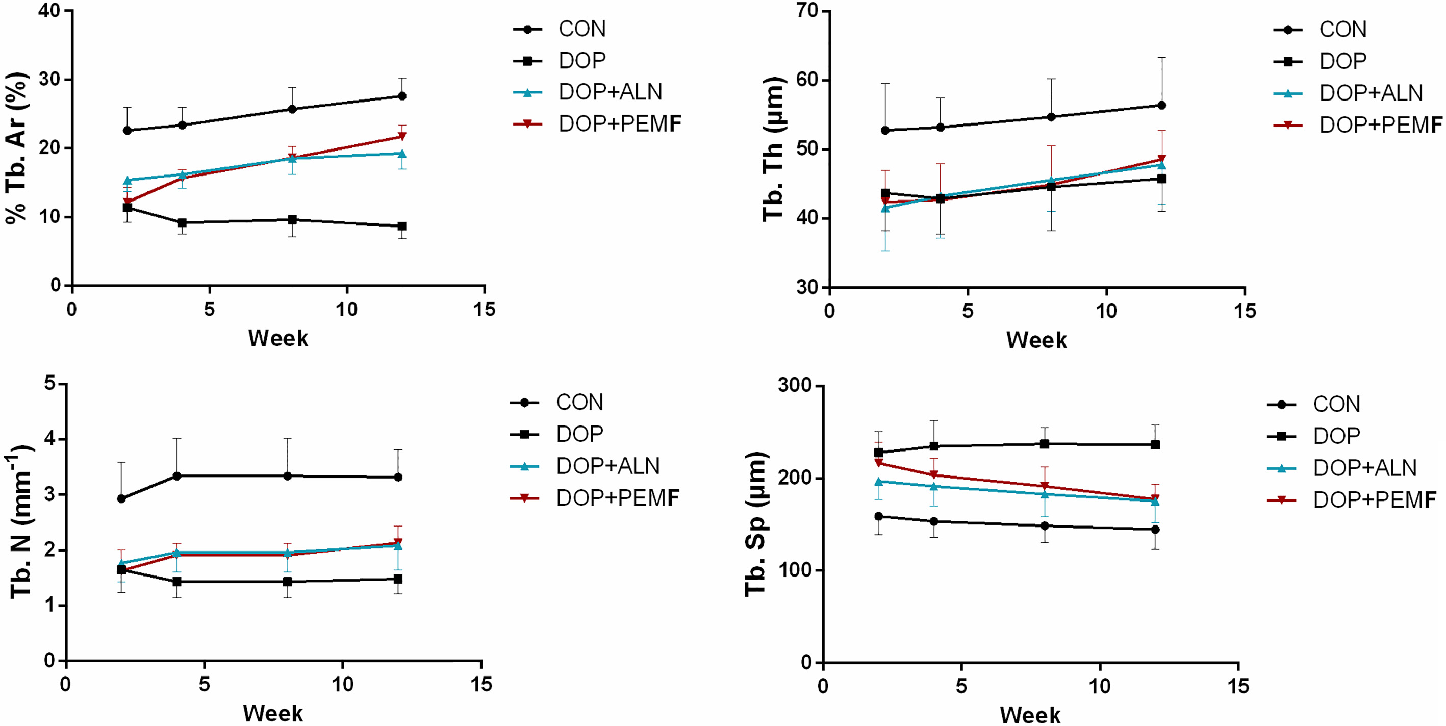
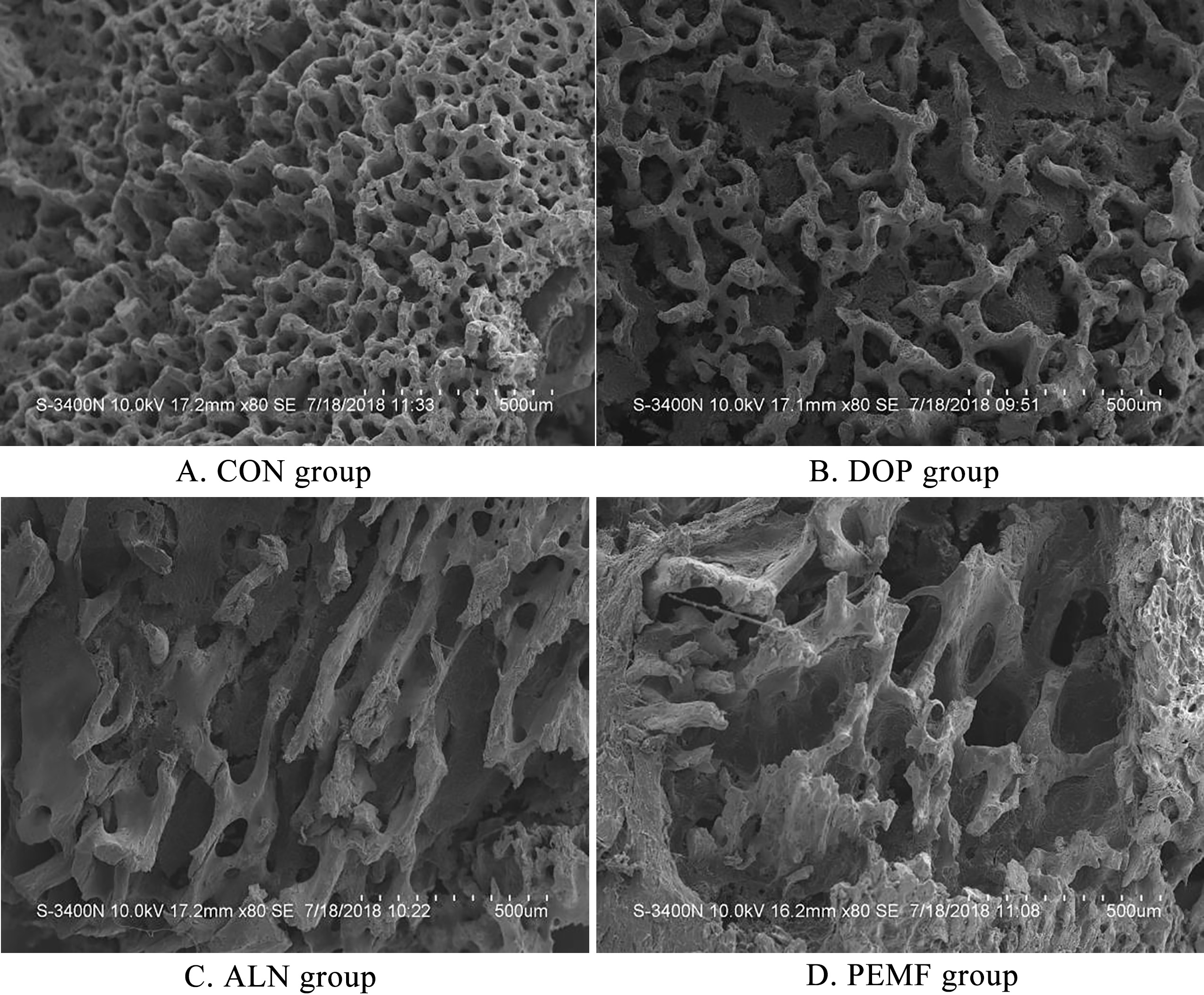
Figure 4.
Morphological changes in bone tissue during PEMF treatment.
3.3PEMF raised serum BGP levels in DOP rats
All groups were found to have higher expressions of BGP in contrast to the CON group. No observable differences were noted between the PEMF and ALN groups at two weeks in comparison to the DOP group. At 4 weeks, the serum concentration of osteocalcin in PEMF group was significantly higher than that in ALN group (
Figure 5.
Serum BGP concentrations changes at each time point.
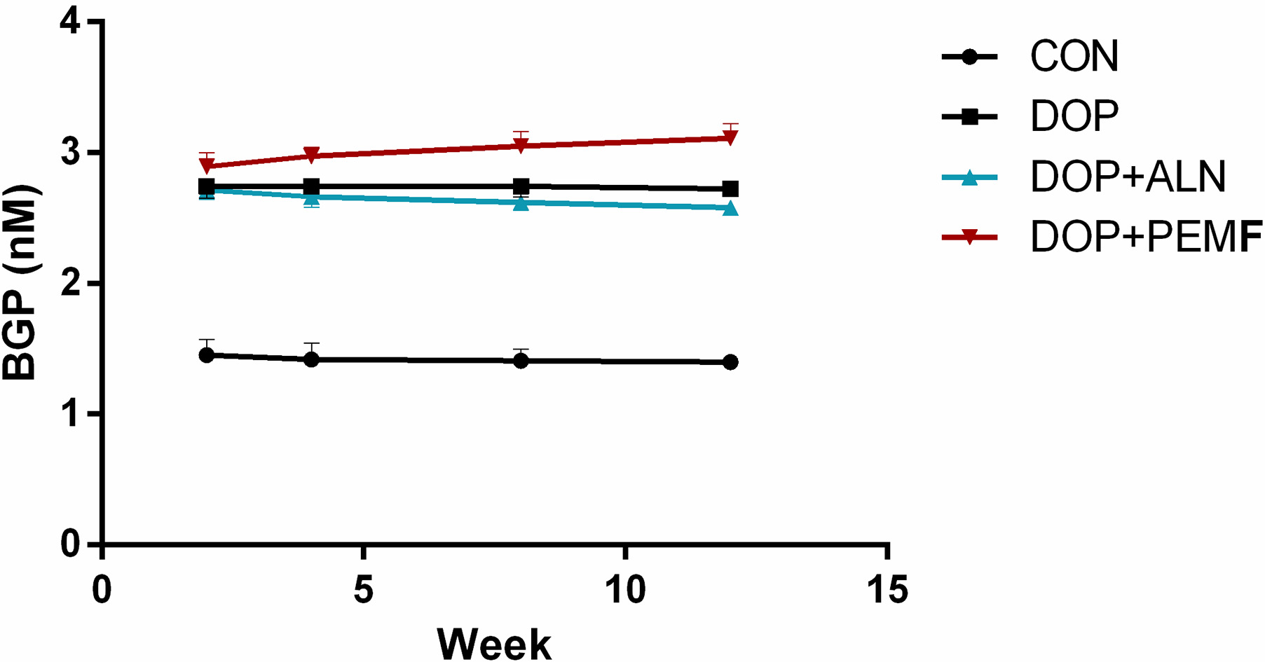
Figure 6.
Serum BMP-2 concentrations in different groups during the treatment period.
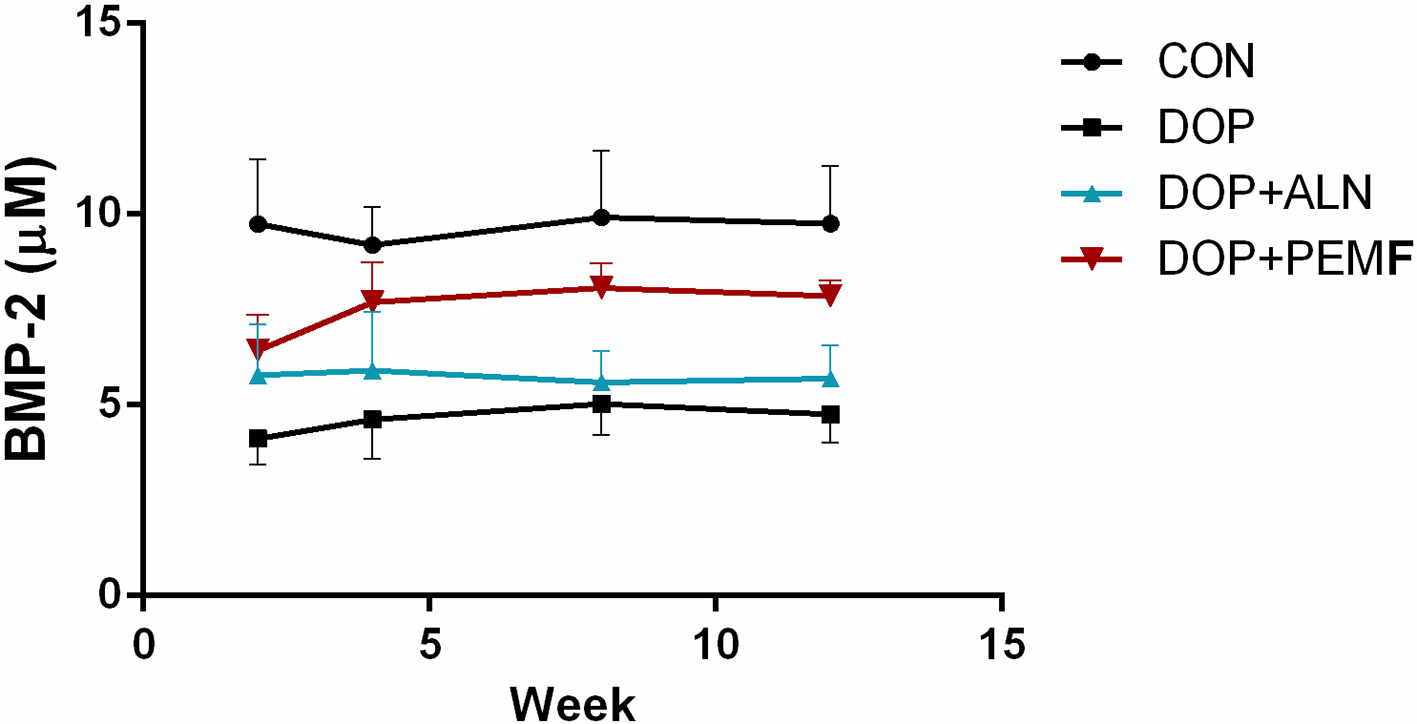
3.4PEMF increased BMP-2 positive expression
The expression of BMP-2 was significantly increased following drug administration in the ALN group in contrast to the CON group (
4.Discussion
The main pathophysiological changes in DOP are bone loss and bone microstructural changes accompanied by accelerated bone growth and slowed formation, leading to a higher fracture risk. DOP treatment is based on enhancement of fracture resistance and reduce the incidence of fractures [5, 6]. Bone loss is not the only indicator of bone strength, as DOP is also associated with alterations in cortical bone thickness and cancellous bone structure [7]. Bone biomechanics is the study of the mechanical characteristics of bone tissue, mechanical parameters under external action, and the biological effects of bone after stress, which is a reliable bone quality evaluation method [8]. Research regarding the mechanism of prevention or treatment of osteoporosis should focus on the effects of biomechanical changes. The geometry of the bone determines the structural mechanical properties, and the fine structure of the bone determines its mechanical properties [9, 10].
Primary therapeutic approaches to DOP focuses on inhibiting bone resorption and improving osteoblastic activity. Several reports have suggested that PEMF is able to promote osteoblast differentiation and proliferation [11, 12]. However, varying magnetic field conditions confer different effects on cellular differentiation and proliferation. According to the conditions reported in the literature [13, 14] combined with self-designed equipment using 3.82 mT and 10 Hz magnetic field conditions, the current research confirms that a 0.15 Gs strength magnetic field could promote bone healing and avoids excessive strength that affects the balance of hormones. In the present study, PEMF effects on bone turnover in disuse osteoporosis were studied by examining bone morphology, bone biomechanical properties, and BMP-2 and BGP concentrations. During the treatment period, Tb-Th values did not vary significantly between the PEMF, ALN and DOP groups, the compensatory thickening of bone trabecula in DOP group may cause such results.
The bone mineral density measurements 2 weeks after hind limb breaking were decreased significantly. Furthermore, the bone mineral density in each model group was decreased in contrast to the CON group (
BGP is a non-collagen protein synthesized and secreted by osteoblasts and is a biological indicator of indirect response to bone metabolism [15]. In the presence of calcium ions, binding to hydroxyapatite to stabilize its conformation is considered the most characteristic marker for osteoblast differentiation [16]. The present data indicates that alendronate may inhibit bone resorption by reducing the functional status of osteoclasts. BGP concentration was significantly increased after modeling in the PEMF group, confirming that PEMF prevents disuse osteoporosis by promoting bone formation.
The most effective factor for osteoinduction is bone morphogenetic proteins [17, 18], which are a family of structurally and functionally related polypeptide factors that induce undifferentiated mesenchymal cells in vitro and in vivo. BMP-2 is the most important bone formation growth factor [19]. BMP-2 has the ability to promote the proliferation and differentiation of mesenchymal stem cells into osteoblasts. Moreover, BMP-2 can be combined with a variety of carriers composed of different types of bioactive repair materials. Following 2 weeks of intervention, the tibia immunohistochemistry index was significantly raised in the ALN group; however, in the PEMF group, the tibia immunohistochemistry index began to increase at 4 weeks, showing that treatment with PEMF was more effective as compared with treatment with alendronate. Later, in the 8th week of treatment, the tibia immunohistochemistry index exceeded that in the ALN group, being much greater by week 12, indicating that PEMF took effect later than alendronate, but the duration of action was longer.
5.Conclusion
PEMF can actively and limitedly prevent and treat disuse osteoporosis, which was achieved by changing the microstructure of bone tissue. BGP and BMP-2 play vital roles in PEMF in improving the treatment of disuse osteoporosis, and PEMF and alendronate enhanced the biomechanical properties of disuse osteoporosis bone. These results show that alendronate confers an immediate effect in preventing and treating disuse osteoporosis, but PEMF offers a less toxic and more reliable outcome in the long term.
Acknowledgments
This study was funded by The First Hospital of Harbin under the High-level Introduction Project (Nos 2013SYYRCYJ05-2 and 2014SYYRCYJ06-3).
Conflict of interest
None to report.
References
[1] | Li, J, Yang, S, Li, X, Liu, D, Wang, Z, Guo, J, et al. Role of endoplasmic reticulum stress in disuse osteoporosis. Bone. (2017) ; 97: : 2-14. doi: 10.1016/j.bone.2016.12.009. |
[2] | Blain, H, Chavassieux, P, Portero-Muzy, N, Bonnel, F, Canovas, F, Chammas, M, et al. Cortical and trabecular bone distribution in the femoral neck in osteoporosis and osteoarthritis. Bone. (2008) ; 43: (5): 862-868. doi: 10.1016/j.bone.2008.07.236. |
[3] | Zofková, I, Nemcikova, P, and Matucha, P. Trace elements and bone health. Clinical Chemistry and Laboratory Medicine. (2013) ; 51: (8): 1555-1561. doi: 10.1515/cclm-2012-0868. |
[4] | Shen, WW, and Zhao, JH. Pulsed electromagnetic fields stimulation affects BMD and local factor production of rats with disuse osteoporosis. Bioelectromagnetics. (2010) ; 31: (2): 113-119. doi: 10.1002/bem.20535. |
[5] | Li, J, Zeng, Z, Zhao, Y, Jing, D, Tang, C, Ding, Y, et al. Effects of low-intensity pulsed electromagnetic fields on bone microarchitecture, mechanical strength and bone turnover in type 2 diabetic db/db mice. Sci Rep. (2017) ; 7: (1): 10834. doi: 10.1038/s41598-017-11090-7. |
[6] | Kasturi, GC, Cifu, DX, and Adler, RA. A review of osteoporosis: Part I. Impact, pathophysiology, diagnosis and unique role of the physiatrist. PM & R: The Journal of Injury, Function, and Rehabilitation. (2009) ; 1: (3): 254-260. doi: 10.1016/j.pmrj.2008.12.005. |
[7] | Smith, EM, Comiskey, CM, and Carroll, AM. A study of bone mineral density in adults with disability. Archives of Physical Medicine and Rehabilitation. (2009) ; 90: (7): 1127-1135. doi: 10.1016/j.apmr.2008.09.578. |
[8] | Mullard, A. FDA rejects first-in-class osteoporosis drug. Nature Reviews. Drug Discovery. (2017) ; 16: (9): 593. doi: 10.1038/nrd.2017.176. |
[9] | Polat, HB, and Beyazal, MS. The effect of cholecystectomy on 25-hydroxyvitamin D levels and bone mineral density in postmenopausal women. Archives of Osteoporosis. (2018) ; 13: (1): 61. doi: 10.1007/s11657-018-0458-0. |
[10] | Bi, X, Grafe, I, Ding, H, Flores, R, Munivez, E, Jiang, MM, et al. Correlations between bone mechanical properties and bone composition parameters in mouse models of dominant and recessive osteogenesis imperfecta and the response to anti-TGF-β treatment. Journal of Bone and Mineral Research: The Official Journal of the American Society for Bone and Mineral Research. (2017) ; 32: (2): 347-359. doi: 10.1002/jbmr.2997. |
[11] | Fini, M, Torricelli, P, Giavaresi, G, Aldini, NN, Cavani, F, Setti, S, et al. Effect of pulsed electromagnetic field stimulation on knee cartilage, subchondral and epyphiseal trabecular bone of aged Dunkin Hartley guinea pigs. Biomedicine & Pharmacotherapy = Biomedecine & Pharmacotherapie. (2008) ; 62: (10): 709-715. doi: 10.1016/j.biopha.2007.03.001. |
[12] | Yamaguchi, DT, Huang, J, Ma, D, and Wang, PK. Inhibition of gap junction intercellular communication by extremely low-frequency electromagnetic fields in osteoblast-like models is dependent on cell differentiation. Journal of Cellular Physiology. (2002) ; 190: (2): 180-188. doi: 10.1002/jcp.10047. |
[13] | Oxlund, BS, Ørtoft, G, Andreassen, TT, and Oxlund, H. Low-intensity, high-frequency vibration appears to prevent the decrease in strength of the femur and tibia associated with ovariectomy of adult rats. Bone. (2003) ; 32: (1): 69-77. |
[14] | Judex, S, Lei, X, Han, D, and Rubin, C. Low-magnitude mechanical signals that stimulate bone formation in the ovariectomized rat are dependent on the applied frequency but not on the strain magnitude. Journal of Biomechanics. (2007) ; 40: (6): 1333-1339. doi: 10.1016/j.jbiomech.2006.05.014. |
[15] | Zhu, X, Luo, J, Chen, X, Wang, J, Wang, G, Li, H, et al. Expression characteristic and significance of interleukin-6, nuclear factor kappa beta, and bone formation markers in rat models of osteoporosis. Translational Research: The Journal of Laboratory and Clinical Medicine. (2008) ; 152: (1): 18-23. doi: 10.1016/j.trsl.2008.05.003. |
[16] | Levy, MM, Joyner, CJ, Virdi, AS, Reed, A, Triffitt, JT, Simpson, AH, et al. Osteoprogenitor cells of mature human skeletal muscle tissue: An in vitro study. Bone. (2001) ; 29: (4): 317-322. |
[17] | Luther, G, Wagner, ER, Zhu, G, Kang, Q, Luo, Q, Lamplot, J, et al. BMP-9 induced osteogenic differentiation of mesenchymal stem cells: Molecular mechanism and therapeutic potential. Current Gene Therapy. (2011) ; 11: (3): 229-240. |
[18] | Jones, AL, Bucholz, RW, Bosse, MJ, Mirza, SK, Lyon, TR, Webb, LX, et al. Recombinant human BMP-2 and allograft compared with autogenous bone graft for reconstruction of diaphyseal tibial fractures with cortical defects. A randomized, controlled trial. The Journal of Bone and Joint Surgery. American Volume. (2006) ; 88: (7): 1431-1441. doi: 10.2106/jbjs.E.00381. |
[19] | Gardner, OF, Fahy, N, Alini, M, and Stoddart, MJ. Differences in human mesenchymal stem cell secretomes during chondrogenic induction. European Cells & Materials. (2016) ; 31: : 221-235. |


