
The impact of rehabilitation in bone loss management of patients with spinal cord injury: A systematic review
Abstract
BACKGROUND:
Spinal cord injury (SCI) is a disabling condition characterized by multilevel skeletal muscle impairment and rapid cortical and trabecular bone loss. Rehabilitation is a cornerstone of the long-term management of patients with SCI; however, the optimal rehabilitation strategy for improving bone health has not been fully characterized.
OBJECTIVE:
To characterize the current evidence supporting different rehabilitation interventions improving bone health in patients with SCI.
METHODS:
On November 17
RESULTS:
Out of 499 records, 11 RCTs met the eligibility criteria and were included. Electrical stimulation combined with physical exercise was assessed by 5 studies, standing intervention was assessed by 3 studies, vibration was assessed by 1 study, ultrasound therapy was assessed by 1 study, and electroacupuncture combined with a pulsed magnetic field was assessed by 1 study. The rehabilitation intervention was administered combined with pharmacological treatment (3 studies) or alone (8 studies). Positive effects in terms of BMD were reported by 3 studies. The quality assessment revealed some concerns in 9 out of 11 studies, in accordance with the Cochrane Risk of Bias assessment – version 2.
CONCLUSION:
Our data suggest that multicomponent interventions including rehabilitation might be considered a suitable option to improve bone health management in SCI patients. Further studies are mandatory to characterize the optimal combination of non-pharmacological interventions reducing bone loss and improving the risk of fractures in patients with SCI.
1.Introduction
Spinal cord injury (SCI) is a disabling neurological condition characterized by the loss of both motor and sensory function and visceral dysfunction [1, 2, 3, 4]. The prevalence of SCI is highly heterogeneous among different countries, ranging from 13.0 per million to 163.4 per million people, with a significant proportion of traumatic spinal cord injuries affecting young adults [5, 6, 7]. Regrettably, SCI is related to extremely high healthcare and assistance costs, while social and personal costs cannot be quantified [5, 6, 7].
After spinal cord injury, bone loss is a common complication related to skeletal muscle system disuse and impaired mechanical stimuli [3, 8]. More in detail, osteoporosis generally affects the skeletal system caudal to the spinal cord damage, with bone loss occurring most rapidly and significantly in the distal femur and proximal tibia [9, 10]. Within two to three years of SCI, individuals show a 50–100% trabecular bone mineral density (BMD) reduction, and 40–80% lower cortical bone mass [11, 12, 13]. As a result, SCI patients have a 20- to 100-fold higher fracture risk compared to the general population [14]. Therefore, it has been proposed that the comprehensive management of bone loss should be performed in patients with SCI, including both pharmacological and non-pharmacological approaches [15].
In this context, growing evidence highlighted the crucial role of a comprehensive rehabilitation program targeting both physical and psychosocial impairment of SCI patients [8, 16, 17, 18, 19, 20]. To date, several studies reported positive effects of physical exercise and physical activity on bone health in several disabling conditions [21]. On the other hand, rehabilitation might effectively target not only bone health but also balance and the risk of falls [22].
Despite these considerations, the optimal rehabilitation strategies to prevent bone loss in people with SCI have yet to be fully characterized [23, 24, 25, 26]. Furthermore, to our knowledge, no previous systematic review including only randomized controlled trials (RCTs) assessed the effects of specific rehabilitation protocols on bone health of SCI patients.
Therefore, the objective of this systematic review was to provide evidence supporting different rehabilitation strategies for improving bone health in patients with SCI. Moreover, we aimed at characterizing the effects of specific rehabilitation prescriptions in bone health in order to pave the way to an evidence-based approach preventing bone loss in people with SCI.
2.Methods
2.1Registration
This systematic review of RCTs follows the Preferred Reporting Items for Systematic Reviews and Meta-analyses (PRISMA) statement [27]. A preliminary search has been performed in the international prospective register of systematic reviews (PROSPERO) for comparable review protocols in progress, without finding similar studies. The systematic review was then submitted to PROSPERO and accepted on 27
2.2Search strategy
The literature search has been performed from onset up to November 17
2.3Selection criteria
In accordance with the PICO model [28], we considered eligible all randomized controlled trials (RCT) satisfying the following criteria:
• P) Participants: adult (aged more than 18 years) male or female patients with spinal cord injury, without restriction in terms of time of injury.
• I) Intervention: all rehabilitation interventions administered as exclusive or integrated therapeutic intervention.
• C) Comparator: any comparator.
• O) Outcome: the primary outcomes were bone macroscopical effects, assessed by: i. Bone Mineral Density (BMD); ii. Bone Mineral Content (BMC); iii. Trabecular Bone Thickness (TBTh) and Trabecular Bone Separation (TBSp); iv. bone strength indices [Compressive Strength Index (CSI), Cortical Thickness Index (CTI), Cortical Bone Volume (CBV), Buckling Ratio (BR), and Torsional Strength Index (TSI)]; v. quantitative bone ultrasound (qUS) parameters; vi. Cross-Sectional Area (CSA) of red and yellow bone marrow and cortical and trabecular bone. The secondary outcomes were: i. changes in body composition; ii. change in bone biomarkers; iii. hormonal and metabolic changes; iv. changes in physical functioning and physical performance.
Only RCTs published in peer-reviewed International Journal were included. No publication date restriction was applied.
The exclusion criteria were: i. participants with pregnancy, clinical instability (defined as hemodynamic alterations, respiratory events, abnormal laboratory values, reduced level of consciousness or temperature alterations), or palliation; ii. studies involving animals; iii. doctorate theses, conference proceedings, and reviews of the literature; iv. not RCT studies; v. language other than English.
Firstly, the articles resulting from the database-specific search strings were examined for duplication removal. Subsequently, the titles and abstracts were screened by two investigators that independently excluded records that did not meet the inclusion criteria. Lastly, the selected articles were examined in full text, and the articles included in the review were thus extracted. Any conflicting record along this chain was discussed between the two investigators, and in case of unresolved disagreement, a third reviewer was involved to reach consensus.
2.4Data extraction and synthesis
All data were extracted by two independent reviewers through Excel database. Any difference was solved by discussion between the two reviewers or by consulting a third reviewer.
The data extracted were: i. authors, journal, publication year, and nationality; ii. study design; iii. sample characteristics [number of participants, mean age and age range, gender, time of injury, lesion level and grade, baseline body mass index (BMI) and osteoporosis grade]; iv. comparator characteristics; v. intervention characteristics [type, duration, frequency, and intensity of treatment in the experimental group and in the control group(s)]; vi. primary and secondary outcomes; vii. duration of the study (with possible follow-up); viii study results.
Data were independently synthesized by two reviewers. A qualitative analysis of the evidence was performed, given that the heterogeneity of the study samples, the type of intervention and the outcome measures did not allow a quantitative analysis of the data.
A subgroup analysis was performed based on participant characteristics, time from SCI, type of intervention, and outcome assessed.
2.5Quality assessment and risk of bias
The qualitative analysis of the studies included in the review was carried out using the PEDro scale [29], which consists of 11 items. In particular, the first item assesses the external validity (or applicability) of the study although is not considered for the purposes of the total score; items 2–9 evaluate the internal validity of the study, while items 10–11 evaluate the interpretability of the results based on the statistical information. Each criterion has “yes” and “no” response options, 1 point for each item whose answer is “yes”, 0 points for items whose answer is “no”. The maximum score obtainable is 10/10. Scores from 9 to 10 were considered “excellent”, 6 to 8 were considered “good”, 4 to 5 were considered “fair”, and
The risk of bias assessment of the studies included in the review was performed following the Cochrane Risk of Bias assessment – version 2 (RoB 2) [30], a validated tool for assessing the risk of bias in RCTs. It includes five different domains, each subdivided in criteria that are judged individually (low, high, unclear risk of bias). The overall judgment of a domain derives from the overall judgment of the various criteria. Studies were considered with low risk of bias if all domains evaluated had low risk of bias. Studies in which at least one domain presented unclear risk, were considered studies with some critical issues. Lastly, studies were considered with high risk of bias if at least one domain had a high risk of bias [30].
3.Results
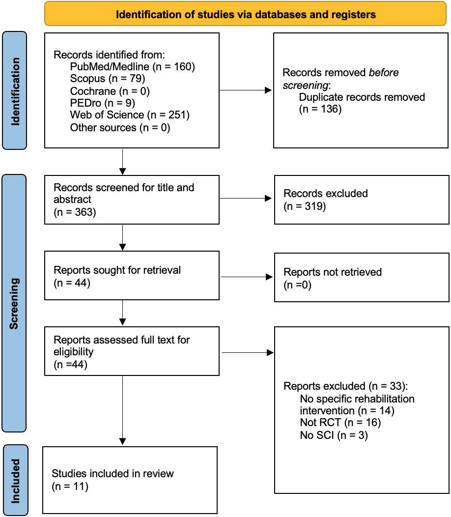
Figure 1.
PRISMA 2020 flow diagram.
3.1Study characteristics
Overall, a total of 499 records resulted from the 5 databases assessed, while no other records were identified from other sources. After duplication removal, a total of 363 studies were assessed for eligibility and screened for title and abstract. As a result, 319 records were excluded, and 44 studies were sought for retrieval and then screened in full text. Lastly, 11 studies [31, 32, 33, 34, 35, 36, 37, 38, 38, 40, 41] were included in the present systematic review. Figure 1 shows the PRISMA flow diagram, reporting further details about the article selection process. Supplementary Table 2 summarizes the reason for exclusion of the remaining 33 studies assessed full-text for eligibility.
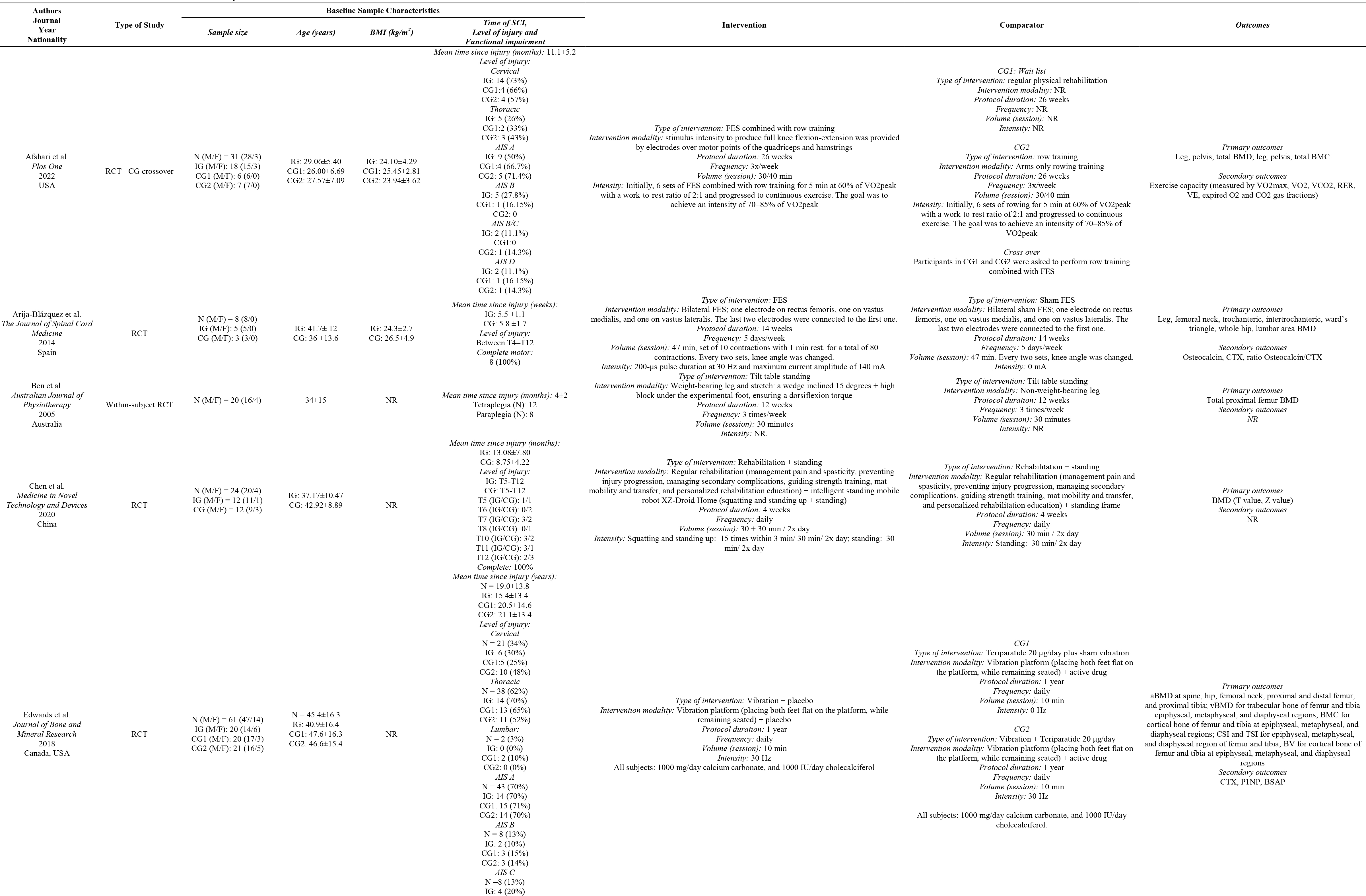
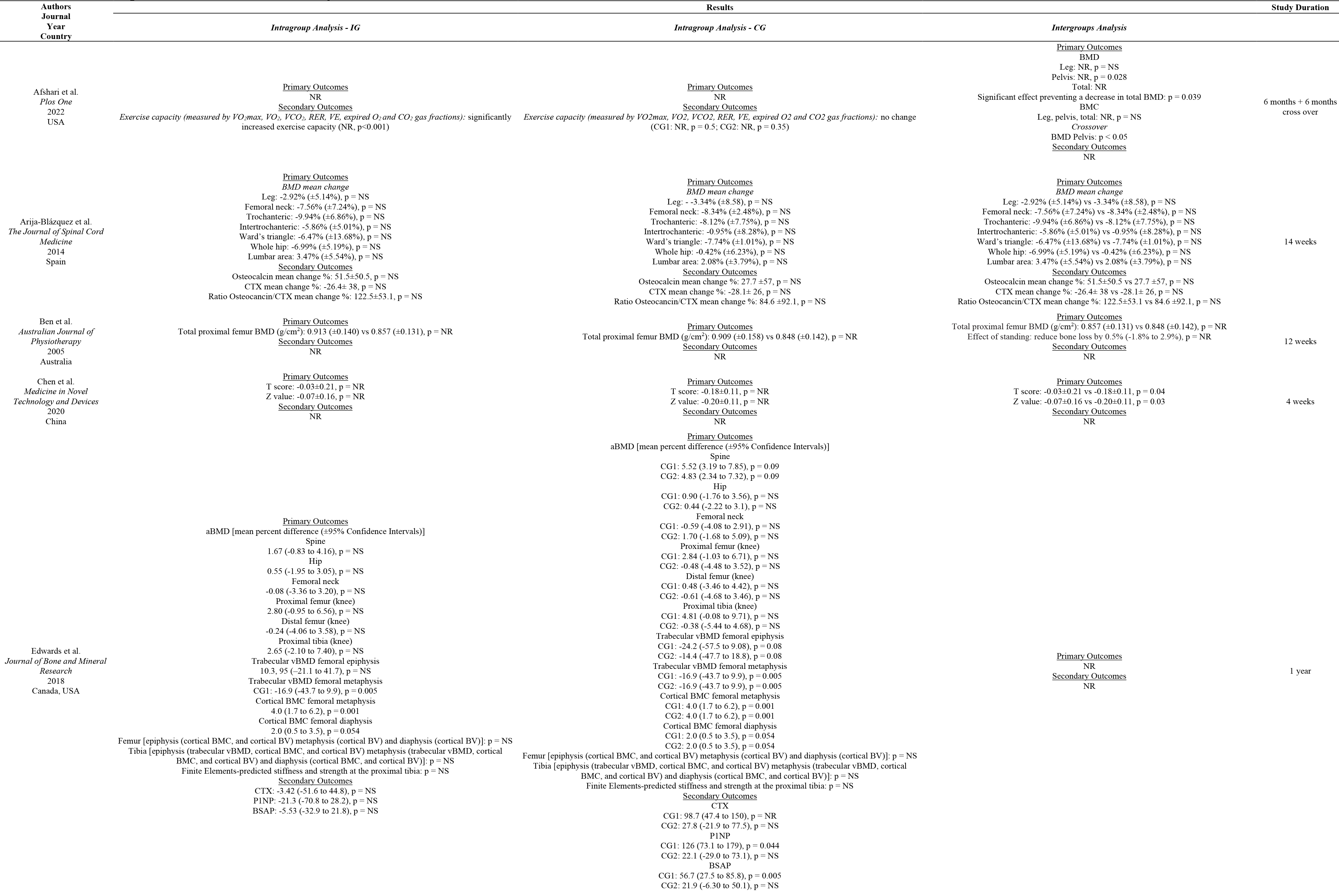
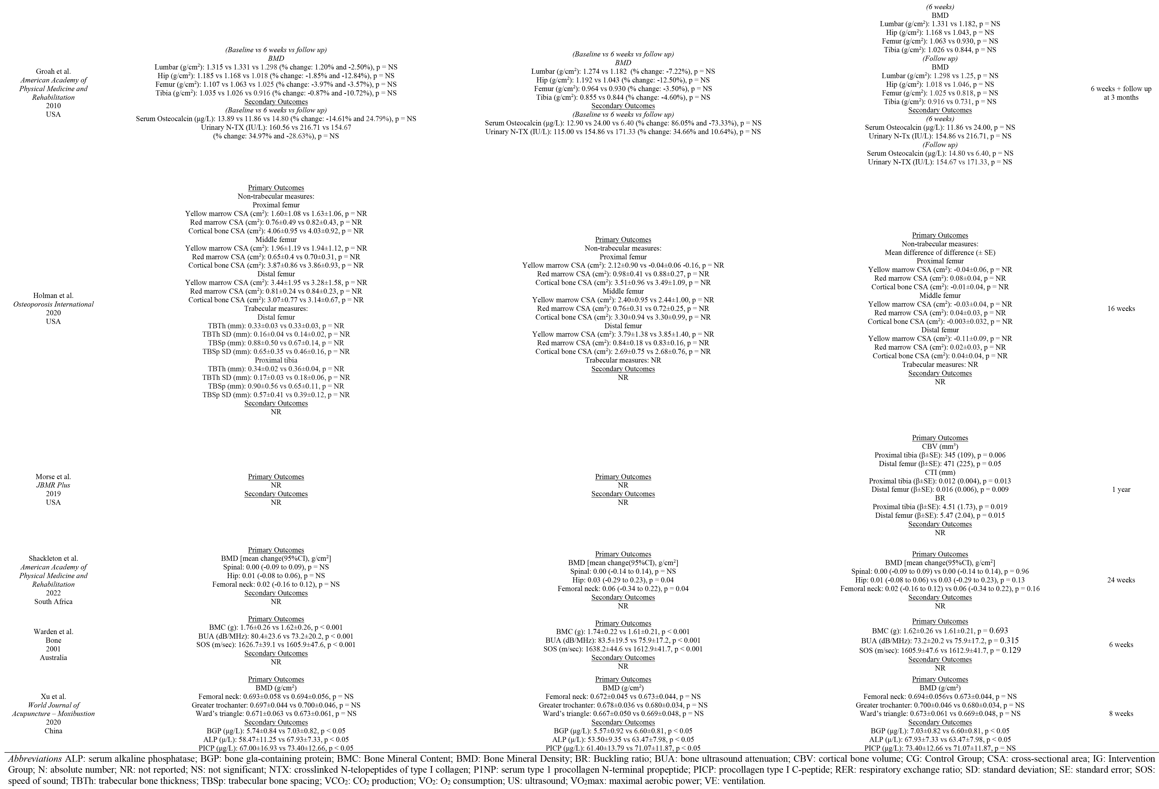
The following studies were included in the present systematic review: Afshari et al. [31], Arija-Blázquez et al. [32], Ben et al. [33], Chen et al. [34], Edwards et al. [35], Groah et al. [36], Holman et al. [37], Morse et al. [38], Shackleton et al. [39], Warden et al. [40], Xu et al. [41].
The studies included were published between2001 [40] and 2022 [31, 39] and were conducted in America (USA
The present review included a total of 301 patients (250 males, and 51 females). The patients were characterized by a mean age ranging between 23.9
Patients with cervical SCI were assessed in five studies [31, 35, 37, 39, 40]; patients with thoracic SCI were evaluated in eight studies [31, 32, 34, 35, 37, 39, 40, 41], and patients with lumbar SCI was assessed in two studies [35, 41]. Two studies did not clarify the level of injury [33, 38]. Asia Impairment Scale (AIS) was the most used tool used to characterize the functional impairment of SCI (
Table 1
Literature search strategy on different databases
| PubMed: (“spinal cord injury” [MeSH Terms] OR “spinal cord trauma” [All Fields] OR “spinal cord injuries” [All Fields] OR “spinal cord transection” [All Fields] OR “spinal cord laceration” [All Fields]) AND (“osteoporosis” [MeSH Terms] OR “osteoporosis” [All Fields] OR “bone loss” [All Fields]) AND (“management” [All Fields] OR “treatment” [All Fields] OR “therapy” [All Fields] OR “therapies” [All Fields] OR “treatments” [All Fields] OR “prevention” [All Fields]) AND (“bone density” [MeSH Terms] OR “bone mineral density” [All Fields] OR “bone mineral content” [All Fields] OR “risk of fracture” [All Fields] OR “risk of fractures” [All Fields]). |
| Physiotherapy Evidence Database (PEDro): spinal cord injur* osteoporosis |
| Web of Science: (spinal cord injury) AND (osteoporosis OR bone loss) AND (management OR treatment OR prevention) AND (bone mineral density OR risk of fracture) |
| Cochrane Central Register of Controlled Trials (CENTRAL) Advanced Search: ID Search #1 MeSH descriptor: [Spinal Cord Injuries] explode all trees #2 MeSH descriptor: [Osteoporosis] explode all trees #3 MeSH descriptor: [Therapeutics] explode all trees #4 MeSH descriptor: [Bone Density] explode all trees #5 #1 AND (#2 AND #3) AND #4 |
| Scopus: TITLE-ABS-KEY: spinal cord injury AND ((osteoporosis OR osteoporosis) AND (treatment OR management OR therapy OR prevention)) AND (bone mineral density OR bmd OR risk of fracture) |
Table 1 summarizes in detail the sample characteristics of both intervention groups and comparator groups of each study included in the present review.
3.2Intervention and control characteristics
Heterogeneous rehabilitation modalities were assessed by the RCTs included. In particular, rehabilitation interventions currently proposed were:
• Functional Electrical Stimulation (FES) combined with physical exercise was assessed in five studies [31, 32, 36, 37, 38]. Afshari et al. [31] positioned electrodes on the quadriceps and hamstrings muscles and delivered a stimulus to produce full knee flexion-extension during arms rowing training. The group compared this intervention against conventional physical rehabilitation arm and rowing training arm [31]. Similarly, Morse et al. [38] assessed the effects of FES and rowing training combined with Zoledronic acid 5 mg/100mL [38]. Arija-Blázquez et al. [32] performed bilateral FES, placing three electrodes on quadriceps muscle to produce contraction, comparing it to sham FES [32]. Groah et al. [36] performed FES bilaterally on the quadriceps muscle combined with non-specified individualized inpatient rehabilitation program, comparing it to the same rehabilitation program without FES [36]. Holman et al. [37] used FES to perform open kinetic chain neuromuscular electrical stimulation-evoked resistance training, with electrodes applied on distal and proximal thigh. The intervention group (IG) was also treated with transdermal testosterone patches, compared with transdermal testosterone patches alone [37].
• Standing modalities were studied in threeRCTs [33, 34, 39]. More in detail, Ben et al. [33] tested weight-bearing and stretch achieved through a block under the experimental foot combined with a tilt table standing. The control was the opposite foot, without weight-bearing [33]. Chen et al. [34] assessed an intelligent standing mobile robot (XZ-Droid Home) with which the patient performed squatting and standing up, and tested it against a regular standing frame. Both IG and control group (CG) underwent rehabilitation, comprising management of pain and spasticity, preventing injury progression, managing secondary complications, guiding strength training, mat mobility and transfer, and personalized rehabilitation education [34]. Lastly, Shackleton et al. [39] used robotic locomotor training to achieve exoskeleton walking in IG. This training modality was compared to activity-based training, which focused on prehabilitation, muscle recruitment, posture and joint stability, resistance and endurance training, pregait, and gait training [39].
• Vibration therapy was studied by Edwards et al. [35]. The group used a vibration platform over which the patient placed both feet flat while remaining seated. This IG was compared to the administration of teriparatide 20
• Ultrasound (US) therapy was assessed by Warden et al. [40]. The authors applied active coupled US-heads on medial and lateral surfaces of the calcaneus, comparing it with sham treatment on contralateral calcaneus [40].
• Electroacupuncture combined with a pulsed magnetic field was assessed by Xu et al. [41]. More in detail, electroacupuncture was performed by treating points GB 34 and GB 39 on both sides. This treatment was combined with XT-20 0 0B Osteoporosis Treatment Apparatus (pulsed magnetic field below the level of spinal cord injury), Reneed tablet, and rehabilitation treatment (standing, limb air pressure, limb function training) [41].
Rehabilitation was combined with pharmacological and nutritional intervention as it follows. Morse et al. [38] treated IG patients with a one-time infusion of Zoledronic acid 5 mg/100mL solution [38]. Holman et al. [37] used transdermal testosterone patches in both IG and CG. In the study by Edwards et al. [35], control groups received teriparatide 20
3.3Primary outcomes
BMD was evaluated in eight studies [31, 32, 33, 34, 35, 36, 39, 41]. Afshari et al. [31] assessed leg, pelvis and total BMD, and reported a significantly lower decrease in pelvis BMD (
BMC was assessed in three studies [31, 35, 40]. Afshari et al. [31] did not find significant differences in leg, pelvis, and total BMC (
Trabecular bone thickness and trabecular bone separation parameters were assessed by Holman et al. [37] for distal femur and proximal tibia, though the authors did not perform statistical inference and multiple comparisons of the results [37].
Bone strength indices were assessed in two studies [35, 38]. Edwards et al. [35] computed CSI and TSI for epiphyseal, metaphyseal, and diaphyseal regions of femur and tibia, without reporting significant differences (
Bone ultrasound attenuation (BUA) and speed of sound (SOS) were evaluated in one study [40]. Although these parameters had a significant worsening over time (
Table 2
Main characteristics of the studies included in the systematic review
|
|
Table 2, continued |
|
Holman et al. [37] assessed the yellow and red bone marrow CSA and cortical bone CSA at proximal, middle, and distal femoral levels, though the authors did not perform statistical inference and multiple comparisons of the results [37]. Further details are shown in Table 2.
3.4Secondary outcomes
Changes in bone biomarkers were assessed in four studies [32, 35, 36, 41].
• Serum alkaline phosphatase (ALP) was analyzed in the study by Xu et al. [41], reporting a significant difference in the intragroup analysis in both groups and in the intergroup analysis (
• Procollagen type I C-peptide (PICP) was analysed in one study [41]. Xu et al. reported a significant difference in the intragroup analysis in both groups (
• Bone gamma-carboxyglutamic acid containing protein (BGP) was analyzed in one study [41], reporting a significant difference in the intragroup analysis and in the intergroup analysis (
• Osteocalcin was evaluated in two studies [32, 36], without reporting significant results (
• Urinary N-terminal telopeptide (N-TX) was evaluated in one study [36], at 6 weeks and at 3 months after intervention without reporting significant differences in intragroup and between group analysis (
• Collagen type 1 cross-linked C-telopeptide (CTX) was analyzed in two studies [32, 35], which did not report significant changes following intervention [32, 35].
• The ratio Osteocancin/CTX was calculated in one study [32], that did not report significant changes [32].
• Type 1 procollagen amino-terminal propeptide(P1NP) was analyzed in one study [35], which did not report significant changes following intervention [35].
• Bone-specific alkaline phosphatase (BSAP) was analyzed in one study [35], which did not report significant changes following intervention [35].
Lastly, changes in physical functioning and physical performance were assessed by Afshari et al. [31], reporting significant improvement in the IG (
Figure 2.
Cochrane Risk of Bias assessment – version 2 (RoB 2).
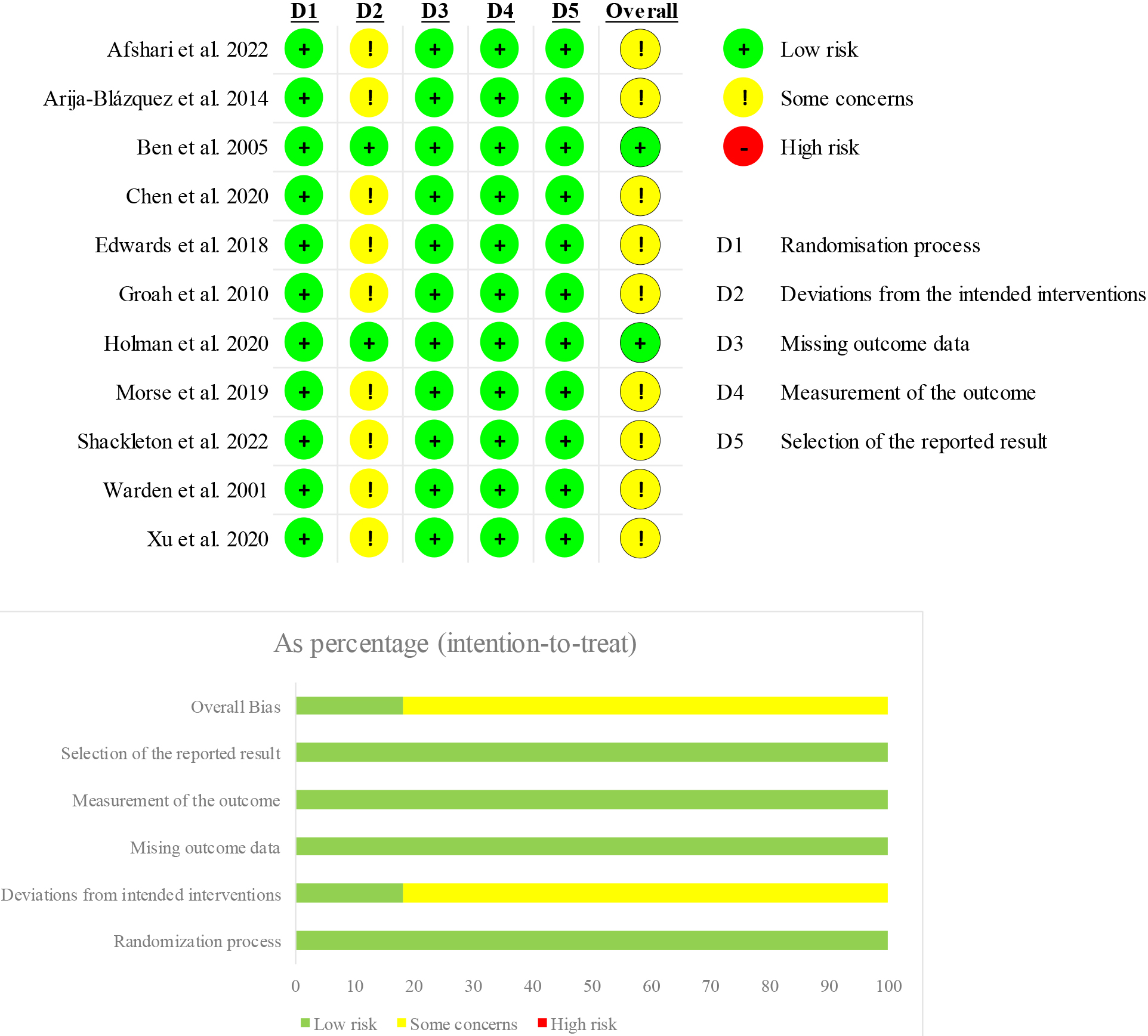
Table 3
Main findings of the studies included in the systematic review
|
|
Table 3, continued |
|
Table 4
Qualitative assessment of the studies included using the PEDro scale
| 1. Eligibility criteria | 2. Random allocation | 3. Concealed allocation | 4. Baseline comparability | 5. Blind subjects | 6. Blind therapists | 7. Blind assessors | 8. Adequate follow-up | 9. Intention- to-treat analysis | 10. Between- group comparisons | 11. Point estimates and variability | Total | |
| Afshari et al. 2022 | 1 | 1 | 0 | 0 | 0 | 0 | 0 | 0 | 0 | 1 | 1 | 3 |
| Arija-Blázquez et al. 2014 | 1 | 1 | 0 | 1 | 1 | 0 | 0 | 1 | 0 | 1 | 1 | 6 |
| Ben et al. 2005 | 1 | 1 | 1 | 1 | 0 | 0 | 1 | 1 | 1 | 1 | 1 | 8 |
| Chen et al. 2020 | 1 | 1 | 0 | 1 | 0 | 0 | 1 | 1 | 0 | 1 | 1 | 6 |
| Edwards et al. 2018 | 1 | 1 | 1 | 1 | 1 | 1 | 0 | 1 | 0 | 1 | 1 | 8 |
| Groah et al. 2010 | 0 | 1 | 0 | 1 | 0 | 0 | 1 | 0 | 0 | 1 | 1 | 5 |
| Holman et al. 2020 | 1 | 1 | 0 | 1 | 0 | 0 | 0 | 1 | 1 | 1 | 1 | 6 |
| Morse et al. 2019 | 1 | 1 | 0 | 0 | 1 | 0 | 0 | 0 | 0 | 1 | 0 | 3 |
| Shackleton et al. 2022 | 0 | 1 | 0 | 1 | 0 | 0 | 0 | 1 | 0 | 1 | 1 | 5 |
| Warden et al. 2001 | 1 | 1 | 0 | 1 | 1 | 0 | 1 | 0 | 0 | 1 | 1 | 6 |
| Xu et al. 2020 | 1 | 1 | 0 | 1 | 0 | 0 | 0 | 1 | 0 | 1 | 1 | 5 |
Note: Eligibility criteria item does not contribute to total score. 0
3.5Quality assessment
In accordance with the PEDro scale [29], two studies resulted in poor quality [31, 38], three studies were characterized as fair [36, 39, 41], while the remaining six studies resulted in good quality scores [32, 33, 34, 35, 37, 40]. Table 3 shows in detail the results of the PEDro scale for each item.
The risk of bias assessment was performed according to the Cochrane Risk of Bias assessment – version 2 (RoB 2) [30]. Some concerns emerged from this analysis in domain 2, specifically “Deviations from the intended interventions”, due to lack of an appropriate analysis estimating the effect of assignment to intervention. This resulted in total outcome of “some concerns” for 9 studies out of 11 [31, 32, 34, 35, 36, 38, 39, 40, 41]. Further details about the risk of bias assessment of each study are shown in Fig. 2.
4.Discussion
Rehabilitation is currently considered a cornerstone in the complex framework of care of SCI patients, with growing research suggesting several therapeutic strategies to improve bone health in these patients [3, 8, 16]. However, bone loss still remains a major issue in SCI patients and the optimal rehabilitation approach to prevent this burdensome condition has not been standardized yet.
Thus, this systematic review of RCTs aimed at summarizing the current evidence supporting the different therapeutic strategies to improve bone health in SCI patients trying to characterize the specific biological effects of a comprehensive rehabilitation approach to these patients.
Interestingly, our findings identified several therapeutic interventions to improve bone health in SCI patients, including FES, physical exercise, vibration therapy, standing, ultrasound, and electroacupuncture combined with pulsed magnetic field. Taken together, the results of the present systematic review underlined that several instrumental therapies were assessed to improve bone health of SCI patients, but with conflicting results and only 3 studies suggested positive effects in terms of BMD improvement at lower limb level [31, 33, 34].
While the effects of physical therapies on bone health are still controversial in the current literature [42, 43, 44], physical exercise is considered a non-pharmacological intervention supported by several guidelines in both osteoporosis prevention and management, due to its multitarget effects on the whole musculoskeletal system, improving both BMD and reducing the risk of falls [45, 46, 47]. Despite these considerations, few studies [31, 32, 36, 37, 38] assessed the effects of physical exercise on SCI patients. However, people with SCI could be affected by detrimental musculoskeletal impairment, with crucial implications in biological responses to physical exercise [48, 49]. Indeed, as part of normal bone metabolism in adult individuals, bone tissue undergoes a continuous process of resorption and formation; when this turnover is at equilibrium, it results in unchanged bone mass, while when there is unbalance, it results in bone changes (growth, aging) and pathology. More in detail, osteoclasts initiate bone resorption, osteoblasts are responsible for bone formation, while osteocytes are the cells dedicated at maintaining bone tissue [50]. On the other hand, as early as 1892, it was determined that mechanical loads in live beings can influence bone architecture. This concept is known as Wolff’s law, which was further explored till defining that loading on load-bearing bones determines the majority of their strength in adult life [51]. In this context, voluntary load bearing is lacking in SCI patients’ life. In this context, it has been suggested that spasticity might have a protective role in bone tissue in patients with neurological disability [52, 53], highlighting the close link between bone and muscle tissues due to their cross-regulation promoted by mechanical forces, in accordance with the mechanostat theory theorized by Frost [54]. On the other hand, it has been proposed that FES treatment might have positive effects on patients affected by spasticity [55]. Despite these considerations, our review underlined that no studies assessed spasticity combined with rehabilitation interventions to manage bone health in SCI patients and more in general the impact of spasticity on bone in these patients is far from being fully characterized.
On the other hand, muscle contraction has been targeted by different rehabilitative programs, including FES and vibration therapy, aiming at increasing mechanical stimuli on the bone and optimizing the protective role of muscle contraction on bone loss in these patients [31, 32, 35, 36, 37, 38].
In accordance with the International Osteoporosis Foundation, non-pharmacological intervention should be considered the first-line therapy in osteoporosis management [56]. However, in patients with a higher risk of fracture, a pharmacological approach is necessary to increase BMD and reduce the risk of fractures [57, 58]. In line with these findings, several studies included in the present review assessed the role of rehabilitation combined with both antiresorptive drugs (zoledronate [38]) and anabolic therapies (teriparatide [35] and testosterone [37]). The recent study by Dionyssiotis et al. [15] reported that level 1 supports intravenous zoledronic acid for preventing sublesional decline, while few studies are currently available about the effects of anabolic treatments for osteoporosis management in this specific population [15].
Lastly, a specific nutritional approach should be part of a comprehensive rehabilitation intervention, given the large consensus about the role of micronutrients in the management of bone health [57]. Despite this evidence, it was surprising to notice that just one study [35] assessed the effects of a combined intervention including rehabilitation and vitamin D
Interestingly, when consulting existing literature, previous reviews assessed the effects of rehabilitation interventions on bone health management of SCI patients. In particular, the review by Sutor et al. [10] assessed the effects of physical exercise and physical activity, providing interesting perspectives about the pathophysiology and molecular mechanisms underpinning physical activity positive effects on bone health of SCI patients. However, several studies with heterogenous designs were included and the authors did not apply a systematic approach. Similarly, the review by Chandrasekaran et al. [59] assessed the effects of FES in patients with SCI. Remarkably, the study highlighted the positive impact of FES in terms of muscle weight, muscle cross-sectional area, and physical functioning. However, the authors did not focus only on bone tissue modifications and the literature was assessed without a systematic approach.
Therefore, to the best of our knowledge, the present study is the first systematic review of RCTs assessing the effectiveness of different rehabilitation strategies to manage bone loss in SCI patients. Altogether, our data highlighted a large gap of knowledge about the potential role of rehabilitation interventions on bone health of SCI patients. On the other hand, a multitarget approach might further implement the synergisms between non-pharmacological and pharmacological interventions, with positive effects not only on bone health but also on functional outcomes of patients with SCI.
Despite these considerations, we are aware that this study has several limitations. In particular, the high heterogeneity of the study interventions limits to draw a quantitative synthesis of the study results. However, this is the first study addressing in a systematic way the effects of rehabilitation interventions on bone health management of SCI patients. In addition, this is the first systematic review assessing the effects of different rehabilitation modalities in this field, despite currently available literature does not allow to compare the effectiveness of different approaches. On the other hand, it should be noted that most of the studies included in the present systematic review showed some concerns in risk of bias assessment, highlighting the need for good quality studies assessing rehabilitation intervention in preventing bone loss of SCI patients.
Lastly, the etiology of SCI lesion, its level, and completeness might severely affect the rehabilitation outcomes, with potential sought of bias in the data synthesis. Therefore, further stratification might improve knowledge about the optimal therapeutic treatment tailored to patient’s characteristics.
5.Conclusions
Altogether, findings of this systematic review underlined that several rehabilitation interventions have been studied to improve bone health in patients with SCI, with promising results reported for physical exercise combined with FES and vibrations. However, several questions are still open about the synergisms of different rehabilitation strategies and the role of a specific rehabilitation approach combined with pharmacological management should still be characterized. Further good-quality studies are needed to improve knowledge about the optimal rehabilitation approach in SCI, improving not only functional outcomes but also bone health of these frail patients.
Ethical approval
Not applicable.
Funding
The authors report no funding.
Informed consent
Not applicable.
Author contributions
Conceptualization, L.L., A.d.S., and M.I.; methodology, L.L., A.d.S., and M.I.; investigation, L.L., A.F., and A.T.; writing – original draft preparation, L.L. and A.F.; writing – review and editing, A.d.S, and M.I.; figures, S.M.; visualization, C.C., S.M, and A.A.; supervision, A.d.S. and M.I. All authors have read and agreed to the published version of the manuscript.
Supplementary data
The supplementary files are available to download from http://dx.doi.org/10.3233//BMR-230006.
Acknowledgments
The authors want to acknowledge Enrico Cavallo for his support of this work.
Conflict of interest
The authors declare that they have no conflict of interest.
References
[1] | Lu X, Battistuzzo CR, Zoghi M, Galea MP. Effects of training on upper limb function after cervical spinal cord injury: a systematic review. Clinical Rehabilitation. (2015) ; 29: (1): 3-13. |
[2] | Stampas A, Tansey KE. Spinal cord injury medicine and rehabilitation. Seminars in Neurology. (2014) ; 34: (5): 524-33. |
[3] | Invernizzi M, de Sire A, Renò F, Cisari C, Runza L, Baricich A, et al. Spinal Cord Injury as a Model of Bone-Muscle Interactions: Therapeutic Implications From in vitro and in vivo Studies. Front Endocrinol (Lausanne). (2020) ; 11: : 204. |
[4] | Invernizzi M, de Sire A, Fusco N. Rethinking the clinical management of volumetric muscle loss in patients with spinal cord injury: Synergy among nutritional supplementation, pharmacotherapy, and rehabilitation. Curr Opin Pharmacol. (2021) ; 57: : 132-9. |
[5] | Pickett GE, Campos-Benitez M, Keller JL, Duggal N. Epidemiology of traumatic spinal cord injury in Canada. Spine. (2006) ; 31: (7): 799-805. |
[6] | Winslow C, Rozovsky J. Effect of spinal cord injury on the respiratory system. American Journal of Physical Medicine & Rehabilitation. (2003) ; 82: (10): 803-14. |
[7] | DeVivo MJ, Krause JS, Lammertse DP. Recent trends in mortality and causes of death among persons with spinal cord injury. Archives of Physical Medicine and Rehabilitation. (1999) ; 80: (11): 1411-9. |
[8] | Invernizzi M, Sire A, Carda S, Venetis K, Renò F, Cisari C, et al. Bone Muscle Crosstalk in Spinal Cord Injuries: Pathophysiology and Implications for Patients’ Quality of Life. Current Osteoporosis Reports. (2020) ; 18: (4): 422-31. |
[9] | Zehnder Y, Lüthi M, Michel D, Knecht H, Perrelet R, Neto I, et al. Long-term changes in bone metabolism, bone mineral density, quantitative ultrasound parameters, and fracture incidence after spinal cord injury: a cross-sectional observational study in 100 paraplegic men. Osteoporosis international: a journal established as result of cooperation between the European Foundation for Osteoporosis and the National Osteoporosis Foundation of the USA. (2004) ; 15: (3): 180-9. |
[10] | Sutor TW, Kura J, Mattingly AJ, Otzel DM, Yarrow JF. The Effects of Exercise and Activity-Based Physical Therapy on Bone after Spinal Cord Injury. International Journal of Molecular Sciences. (2022) ; 23: (2): 608. |
[11] | Eser P, Frotzler A, Zehnder Y, Wick L, Knecht H, Denoth J, et al. Relationship between the duration of paralysis and bone structure: a pQCT study of spinal cord injured individuals. Bone. (2004) ; 34: (5): 869-80. |
[12] | Frotzler A, Berger M, Knecht H, Eser P. Bone steady-state is established at reduced bone strength after spinal cord injury: a longitudinal study using peripheral quantitative computed tomography (pQCT). Bone. (2008) ; 43: (3): 549-55. |
[13] | Edwards WB, Simonian N, Troy KL, Schnitzer TJ. Reduction in torsional stiffness and strength at the proximal tibia as a function of time since spinal cord injury. Journal of Bone and Mineral Research. (2015) ; 30: (8): 1422-30. |
[14] | Frisbie JH. Fractures after myelopathy: the risk quantified. The Journal of Spinal Cord Medicine. (1997) ; 20: (1): 66-9. |
[15] | Dionyssiotis Y, Kalke YB, Frotzler A, Moosburger J, Trovas G, Kaskani E, et al. S1 Guidelines on Bone Impairment in Spinal Cord Injury. J Clin Densitom. (2021) ; 24: (3): 490-501. |
[16] | Morone G, Sire A, Cinnera A, Paci M, Perrero L, Invernizzi M, et al. Upper Limb Robotic Rehabilitation for Patients with Cervical Spinal Cord Injury: A Comprehensive Review. Brain Sciences. (2021) ; 11: (12): 1630. |
[17] | Invernizzi M, Carda S, Rizzi M, Grana E, Squarzanti DF, Cisari C, et al. Evaluation of serum myostatin and sclerostin levels in chronic spinal cord injured patients. Spinal Cord. (2015) ; 53: (8): 615-20. |
[18] | Solomon RM, Dhakal R, Halpin SJ, Hariharan R, O’Connor RJ, Allsop M, et al. Telerehabilitation for individuals with spinal cord injury in low-and middle-income countries: a systematic review of the literature. Spinal Cord. (2022) ; 60: (5): 395-403. |
[19] | Schultz KR, Mona LR, Cameron RP. Mental Health and Spinal Cord Injury: Clinical Considerations for Rehabilitation Providers. Current Physical Medicine and Rehabilitation Reports. (2022) . |
[20] | de Melo-Neto JS, de Campos Gomes F, de Morais DF, Tognola WA. Spinal cord injury in elderly patients admitted to a tertiary hospital. J Back Musculoskelet Rehabil. (2017) ; 30: (4): 929-36. |
[21] | de Sire A, Lippi L, Aprile V, Calafiore D, Folli A, D’Abrosca F, et al. Pharmacological, Nutritional, and Rehabilitative Interventions to Improve the Complex Management of Osteoporosis in Patients with Chronic Obstructive Pulmonary Disease: A Narrative Review. Journal of Personalized Medicine. (2022) ; 12: (10): 1626. |
[22] | Yeung SSY, Reijnierse EM, Pham VK, Trappenburg MC, Lim WK, Meskers CGM, et al. Sarcopenia and its association with falls and fractures in older adults: A systematic review and meta-analysis. Journal of Cachexia, Sarcopenia and Muscle. (2019) ; 10: (3): 485-500. |
[23] | Lippi L, de Sire A, D’Abrosca F, Polla B, Marotta N, Castello LM, et al. Efficacy of Physiotherapy Interventions on Weaning in Mechanically Ventilated Critically Ill Patients: A Systematic Review and Meta-Analysis. Front Med (Lausanne). (2022) ; 9: : 889218. |
[24] | Bernardes Neto SCG, Torres-Castro R, Lima Í, Resqueti VR, Fregonezi GAF. Weaning from mechanical ventilation in people with neuromuscular disease: a systematic review. BMJ Open. (2021) ; 11: (9): e047449. |
[25] | Yuan X, Lu X, Chao Y, Beck J, Sinderby C, Xie J, et al. Neurally adjusted ventilatory assist as a weaning mode for adults with invasive mechanical ventilation: a systematic review and meta-analysis. Critical Care. (2021) ; 25: (1). |
[26] | Leboff MS, Greenspan SL, Insogna KL, Lewiecki EM, Saag KG, Singer AJ, et al. The clinician’s guide to prevention and treatment of osteoporosis. Osteoporosis International. (2022) ; 33: (10): 2049-102. |
[27] | Page MJ, McKenzie JE, Bossuyt PM, Boutron I, Hoffmann TC, Mulrow CD, et al. The PRISMA 2020 statement: an updated guideline for reporting systematic reviews. BMJ. (2021) ; 372: : n71. |
[28] | Huang X, Lin J, Demner-Fushman D. Evaluation of PICO as a knowledge representation for clinical questions. AMIA Annu Symp Proc. (2006) ; 359-63. |
[29] | Cashin AG, McAuley JH. Clinimetrics: Physiotherapy Evidence Database (PEDro) Scale. J Physiother. (2020) ; 66: (1): 59. |
[30] | Sterne JAC, Savović J, Page MJ, Elbers RG, Blencowe NS, Boutron I, et al. RoB 2: a revised tool for assessing risk of bias in randomised trials. BMJ. (2019) ; 366: : l4898. |
[31] | Afshari K, Ozturk ED, Yates B, Picard G, Taylor JA. Effect of hybrid FES exercise on body composition during the sub-acute phase of spinal cord injury. Plos One. (2022) ; 17: (1). |
[32] | Arija-Blázquez A, Ceruelo-Abajo S, Díaz-Merino MS, Godino-Durán JA, Martínez-Dhier L, Martin JL, et al. Effects of electromyostimulation on muscle and bone in men with acute traumatic spinal cord injury: A randomized clinical trial. J Spinal Cord Med. (2014) ; 37: (3): 299-309. |
[33] | Ben M, Harvey L, Denis S, Glinsky J, Goehl G, Chee S, et al. Does 12 weeks of regular standing prevent loss of ankle mobility and bone mineral density in people with recent spinal cord injuries? Australian Journal of Physiotherapy. (2005) ; 51: (4): 251-6. |
[34] | Chen J, Jin Z, Yao J, Wang H, Li Y, Ouyang Z, et al. Influence of the intelligent standing mobile robot on lower extremity physiology of complete spinal cord injury patients. Medicine in Novel Technology and Devices (2020) Sep; 7: : 100045. |
[35] | Edwards WB, Simonian N, Haider IT, Anschel AS, Chen D, Gordon KE, et al. Effects of Teriparatide and Vibration on Bone Mass and Bone Strength in People with Bone Loss and Spinal Cord Injury: A Randomized, Controlled Trial. J Bone Miner Res. (2018) ; 33: (10): 1729-40. |
[36] | Groah SL, Lichy AM, Libin AV, Ljungberg I. Intensive electrical stimulation attenuates femoral bone loss in acute spinal cord injury. PM R. (2010) ; 2: (12): 1080-7. |
[37] | Holman ME, Chang G, Ghatas MP, Saha PK, Zhang X, Khan MR, et al. Bone and non-contractile soft tissue changes following open kinetic chain resistance training and testosterone treatment in spinal cord injury: an exploratory study. Osteoporos Int. (2021) ; 32: (7): 1321-32. |
[38] | Morse LR, Troy KL, Fang Y, Nguyen N, Battaglino R, Goldstein RF, et al. Combination Therapy With Zoledronic Acid and FES-Row Training Mitigates Bone Loss in Paralyzed Legs: Results of a Randomized Comparative Clinical Trial. JBMR Plus. (2019) ; 3: (5). |
[39] | Shackleton C, Evans R, West S, Derman W, Albertus Y. Robotic Walking to Mitigate Bone Mineral Density Decline and Adverse Body Composition in Individuals With Incomplete Spinal Cord Injury A Pilot Randomized Clinical Trial. American Journal of Physical Medicine & Rehabilitation. (2022) ; 101: (10): 931-6. |
[40] | Warden SJ, Bennell KL, Matthews B, Brown DJ, McMeeken JM, Wark JD. Efficacy of low-intensity pulsed ultrasound in the prevention of osteoporosis following spinal cord injury. Bone. (2001) ; 29: (5): 431-6. |
[41] | Xu ZJ, Xiang L, Qi W, Pan K, Li W, Tan Y, et al. Effect of electroacupuncture combined with osteoporosis treatment apparatus on bone metabolism in patients with spinal cord injury. World Journal of Acupuncture-Moxibustion. (2020) ; 30: (4): 251-5. |
[42] | Lang S, Ma J, Gong S, Wang Y, Dong B, Ma X. Pulse Electromagnetic Field for Treating Postmenopausal Osteoporosis: A Systematic Review and Meta-Analysis of Randomized Controlled Trials. Bioelectromagnetics. (2022) ; 43: (6): 381-93. |
[43] | Zhu S, Li Y, Wang L, Huang J, Song K, Gan X, et al. Pulsed Electromagnetic Fields May Be Effective for the Management of Primary Osteoporosis: A Systematic Review and Meta-Analysis. IEEE Transactions on Neural Systems and Rehabilitation Engineering. (2022) ; 30: : 321-8. |
[44] | de Oliveira RDJ, de Oliveira RG, de Oliveira LC, Santos-Filho SD, Sá-Caputo DC, Bernardo-Filho M. Effectiveness of whole-body vibration on bone mineral density in postmenopausal women: a systematic review and meta-analysis of randomized controlled trials. Osteoporosis international: a journal established as result of cooperation between the European Foundation for Osteoporosis and the National Osteoporosis Foundation of the USA. (2022) . |
[45] | Moayyeri A. The association between physical activity and osteoporotic fractures: a review of the evidence and implications for future research. Ann Epidemiol. (2008) ; 18: (11): 827-35. |
[46] | de Kam D, Smulders E, Weerdesteyn V, Smits-Engelsman BC. Exercise interventions to reduce fall-related fractures and their risk factors in individuals with low bone density: a systematic review of randomized controlled trials. Osteoporosis International: A Journal Established as Result of Cooperation between the European Foundation for Osteoporosis and the National Osteoporosis Foundation of the USA. (2009) ; 20: (12): 2111-25. |
[47] | Suominen H. Muscle training for bone strength. Aging Clin Exp Res. (2006) ; 18: (2): 85-93. |
[48] | Basso DM, Hansen CN. Biological basis of exercise-based treatments: spinal cord injury. PM&R. (2011) ; 3: (6): S73-S7. |
[49] | Neefkes-Zonneveld CR, Bakkum AJ, Bishop NC, van Tulder MW, Janssen TW. Effect of long-term physical activity and acute exercise on markers of systemic inflammation in persons with chronic spinal cord injury: a systematic review. Archives of Physical Medicine and Rehabilitation. (2015) ; 96: (1): 30-42. |
[50] | Seibel MJ. Biochemical markers of bone turnover: part I: biochemistry and variability. Clin Biochem Rev. (2005) ; 26: (4): 97-122. |
[51] | Frost HM. A 2003 update of bone physiology and Wolff’s Law for clinicians. Angle Orthod. (2004) ; 74: (1): 3-15. |
[52] | Garland DE, Stewart CA, Adkins RH, Hu SS, Rosen C, Liotta FJ, et al. Osteoporosis after spinal cord injury. Journal of Orthopaedic Research. (1992) ; 10: (3): 371-8. |
[53] | Eser P, Frotzler A, Zehnder Y, Schiessl H, Denoth J. Assessment of anthropometric, systemic, and lifestyle factors influencing bone status in the legs of spinal cord injured individuals. Osteoporosis International. (2005) ; 16: (1): 26-34. |
[54] | Frost HM. From Wolff’s law to the Utah paradigm: insights about bone physiology and its clinical applications. The Anatomical Record: An Official Publication of the American Association of Anatomists. (2001) ; 262: (4): 398-419. |
[55] | Sivaramakrishnan A, Solomon JM, Manikandan N. Comparison of transcutaneous electrical nerve stimulation (TENS) and functional electrical stimulation (FES) for spasticity in spinal cord injury-A pilot randomized cross-over trial. The Journal of Spinal Cord Medicine. (2018) ; 41: (4): 397-406. |
[56] | International Osteoporosis Foundation (IOF) exercise recommendations. iof bone health; [internet]. available from: https://www.iofbonehealth.org/exercise-recommendations. |
[57] | Kanis JA, Cooper C, Rizzoli R, Reginster J-Y. Review of the guideline of the American College of Physicians on the treatment of osteoporosis. Osteoporosis International. (2018) ; 29: (7): 1505-10. |
[58] | Rossini M, Adami S, Bertoldo F, Diacinti D, Gatti D, Giannini S, et al. Guidelines for the diagnosis, prevention and management of osteoporosis. Reumatismo. (2016) ; 68: (1): 1-39. |
[59] | Chandrasekaran S, Davis J, Bersch I, Goldberg G, Gorgey AS. Electrical stimulation and denervated muscles after spinal cord injury. Neural Regen Res. (2020) ; 15: (8): 1397-407. |


