
Serum aspartate aminotransferase, a novel potential biomarker of prognosis in extranodal natural killer/T cell lymphoma, nasal type
Abstract
BACKGROUND:
Aspartate aminotransferase (AST), an indicator of liver cell damage, was related to the prognosis of certain malignant tumors.
OBJECTIVE:
This study examined the predictive value of AST in patients with extranodal natural killer/T cell lymphoma (ENKTL).
METHODS:
We reviewed 183 cases diagnosed with ENKTL and selected 26 U/L as the optimum cut-off value of AST. We used the univariate and multivariate Cox regression to compare the different AST groups’ overall survival (OS) and progression-free survival (PFS).
RESULTS:
Prior to propensity score matching (PSM), Kaplan-Meier analysis showed that patients in the low AST subgroup had better OS and PFS than the high AST subgroup. Multivariate analysis revealed that AST was an independent indicator for prognosis. After PSM, the low AST subgroup maintained a significantly better OS and PFS than the high AST subgroup.
CONCLUSION:
AST might represent a significant prognostic marker for ENKTL patients.
1.Introduction
Extranodal natural killer T-cell lymphoma, nasal-type (ENKTL), is a rare subtype of non-Hodgkin lymphoma (NHL) with a dismal prognosis [1]. This disease is diversely ethnic and geographic as the incidence is high in Latin America and East Asia [2]. The overall survival of ENKTL has been prolonged by more efficacious treatment strategies; however, their prognosis remains poor [3, 4].
Several predictive models in non-Hodgkin’s lymphoma subtypes remain controversial for ENKTL patients [5, 6]. Some studies optimized the Korean Prognostic Index (KPI) system to present preferable prediction ability by integrating laboratory data [7, 8, 9]. Additionally, some scoring systems have been successively explored for better risk stratifications [10, 11, 12]. Kim et al. proposed the prognostic index (PINK) for ENKTL patients who received L-asparaginase-based regimens. [13]. However, some drawbacks still exist, identification of other validated prognostic markers is essential.
Aspartate aminotransferase (AST), an enzyme with a high level in the liver, was generally tested for liver damage. However, many studies suggest that the AST level is associated with non-liver-related mortality [14, 15, 16, 17]. Tumor cells also produce AST, and the level of AST also correlates with the prognosis of hepatocellular and renal cell carcinoma, breast cancer, and multiple myeloma and high levels of AST indicate poor prognosis [18, 19, 20, 21, 22]. However, few studies on the relationship between AST and lymphoma survival have been illustrated [23]. To our best knowledge, this retrospective study first explored AST’s predictive role in ENKTL patients.
2.Methods
2.1Patient collection
In our study, we collected medical records of 183 eligible ENKTL patients from Shanxi Cancer Hospital from January 2002 to December 2018. The inclusion criteria were as follows: (i) confirmed with ENKTL by both pathological diagnosis and immunohistochemistry, (ii) no previous anticancer treatment, (iii) with adequate clinical and follow-up data. Based on the primary tumor site, ENKTL was classified as upper aerodigestive tract NK/T-cell lymphoma (UENKTL) or extra-UENKTL (EUENKTL) [24].
2.2Data collection
We collected pretreatment data regarding laboratory examinations, age, sex, ECOG score, serum LDH level, systemic B symptoms, Extranodal invasion sites, regional lymph node involvement, Ann Arbor Staging, and the biochemical profile from the electronic medical record system. We obtained the AST value (U/L) from the hospital laboratory database.
Additionally, we also analyzed IPI (age, performance status, stage, LDH level, and extranodal sites) [25], KPI (stage, LDH level, B symptoms, and regional lymph nodes) [26], PINK (age, Ann Arbor stage, distant lymph-node involvement, and non-nasal type disease) [13] and NRI (age, ECOG Performance Status, Ann Arbor stage, LDH level, and PTI) [27] calculated at diagnosis.
2.3Statistical analysis
The primary endpoints were progression-free survival (PFS) and overall survival time ( OS). We calculated the optimal cut-off value for AST by the change point method (SurvMisc package, R project, version 3.6.1) [28]. According to this value, patients were stratified into high AST and low AST groups. We used the Kaplan-Meier method and log-rank test to show the differences in the survival curves and univariate and multivariate analyses to assess the prognostic factors with hazard ratios recorded with 95% confidence intervals. The regression was verified using the 10-fold cross-validation with a seed number of 2022. We considered
Table 1
Clinicopathological features of 183 patients according to the AST
| Characteristics | Number of patients (%) | AST | AST | |
|---|---|---|---|---|
| Age | 0.318 | |||
| | 147(80.33%) | 87(82.86%) | 60(76.92%) | |
| | 36(19.67%) | 18(17.14%) | 18(23.08%) | |
| Sex | 0.040 | |||
| Male | 144(78.69%) | 77(73.33%) | 67(85.90%) | |
| Female | 39(21.31%) | 28(26.67%) | 11(14.10%) | |
| ECOG score | 0.169 | |||
| 0–1 | 147(80.33%) | 88(83.81%) | 59(75.64%) | |
| | 36(19.67%) | 17(16.19%) | 19(24.36%) | |
| Ann Arbor Stage | 0.271 | |||
| I–II | 141(77.05%) | 84(80.00%) | 57(73.08%) | |
| III–IV | 42(22.95%) | 21(20.00%) | 21(26.92%) | |
| B symptoms | 0.032 | |||
| No | 117(63.93%) | 74(70.48%) | 43(55.13%) | |
| Yes | 66(36.07%) | 31(29.52%) | 35(44.87%) | |
| Extranodal sites of involvement | 0.146 | |||
| | 158(86.34%) | 94(89.52%) | 64(82.05%) | |
| | 25(13.66%) | 11(10.48%) | 14(17.95%) | |
| Regional lymph node involvement | 0.002 | |||
| Yes | 66(36.07%) | 28(26.67%) | 38(48.72%) | |
| No | 117(63.93%) | 77(73.33%) | 40(51.28%) | |
| Subtype | 0.269 | |||
| UNKTL | 169(92.35%) | 95(90.48%) | 74(94.87%) | |
| EUNKTL | 14(7.65%) | 10(9.52%) | 4(5.13%) | |
| Serum LDH | ||||
| | 129(70.49%) | 96(91.43%) | 33(42.31%) | |
| | 54(29.51%) | 9(8.57%) | 45(57.69%) | |
| Hemoglobin | 0.566 | |||
| | 43(23.50%) | 23(21.90%) | 20(25.64%) | |
| | 140(76.50%) | 82(78.10%) | 58(74.36%) | |
| ALT | ||||
| | 100(54.64%) | 84(80.00%) | 16(20.51%) | |
| | 83(45.36%) | 21(20.00%) | 62(79.49%) | |
| AST/ALT | ||||
| Low | 111(60.66%) | 83(79.05%) | 28(35.90%) | |
| High | 72(39.34%) | 22(20.95%) | 50(64.10%) | |
| IPI | 0.001 | |||
| 0–1 | 132(72.13%) | 86(81.90%) | 46(58.97%) | |
| 2–5 | 51(27.87%) | 19(18.10%) | 32(41.03%) | |
| KPI | ||||
| 0–1 | 116(63.39%) | 83(79.05%) | 33(42.31%) | |
| 2–4 | 67(36.61%) | 22(20.95%) | 45(57.69%) | |
| PINK | 0.897 | |||
| 0–1 | 147(80.33%) | 84(80.00%) | 63(80.77%) | |
| 2–4 | 36(19.67%) | 21(20.00%) | 15(19.23%) | |
| NRI | ||||
| 0–1 | 86(46.99%) | 63(60.00%) | 23(29.49%) | |
| 2–6 | 97(53.01%) | 42(40.00%) | 55(70.51%) | |
| RT | 0.526 | |||
| No | 61(33.33%) | 33(31.43%) | 28(35.90%) | |
| Yes | 122(66.67%) | 72(68.57%) | 50(64.10%) | |
| L-Asp | 0.189 | |||
| No | 100(54.64%) | 53(50.48%) | 47(60.26%) | |
| Yes | 83(45.36%) | 52(49.52%) | 31(39.74%) | |
| Liver disease | 0.316 | |||
| No | 174(95.08%) | 102(93.60%) | 72(97.30%) | |
| Yes | 9(4.92%) | 7(6.40%) | 2(2.70%) | |
| SCT | 1.000 | |||
| No | 179(97.81%) | 107(98.20%) | 72(97.30%) | |
| Yes | 4(2.19%) | 2(1.80%) | 2(2.70%) |
Abbreviation: LDH, lactate dehydrogenase; IPI, International Prognostic Index; KPI, Korean Prognostic Index; PINK, Prognostic index of natural killer lymphoma; NRI, nomogram-revised risk index; RT, radiotherapy; L-Asp, L-Asparaginase; SCT, allogeneic hematopoietic stem cell transplantation.
2.4Ethical approval
The study was approved by the ethics committees at Shanxi Province Cancer Hospital, Shanxi Hospital Affiliated to Cancer Hospital, Chinese Academy of Medical Sciences, Cancer Hospital Affiliated to Shanxi Medical University and the review board approved to waive the requirement for informed consent. (Number: 2019091). Data collection was carried out via electronic medical records and entered in an anonymized databank. All analyses were performed in accordance with the relevant guidelines and regulations.
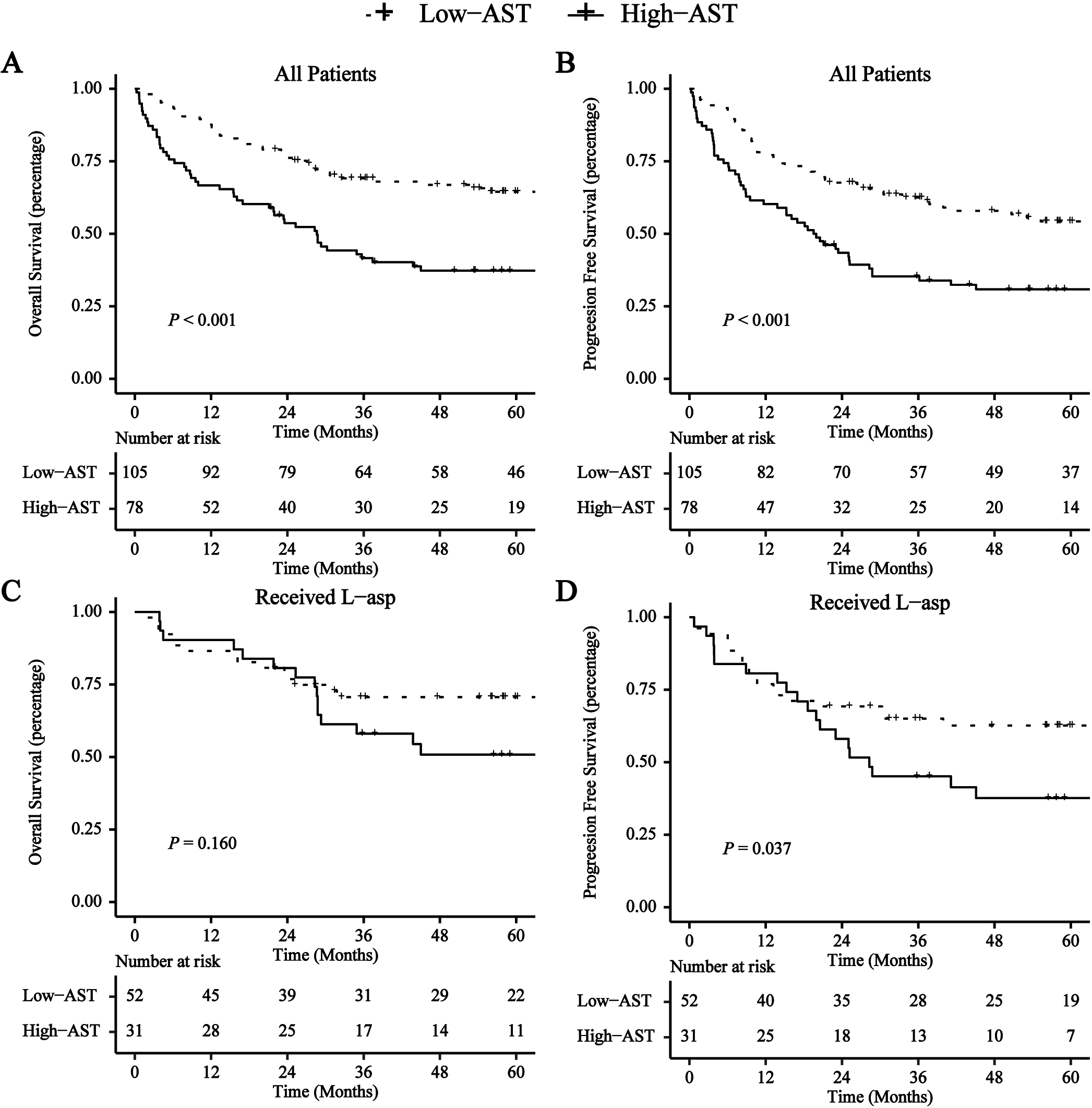
Figure 1.
Survival curves for OS and PFS based on AST (
3.Results
3.1Patients’ characteristics
Table 1 shows the clinical parameters of the patients. We obtained AST before treatment. At the time of diagnosis, the median AST was 24 U/L, and the most discriminative cut-off value was 26 U/L. We compared baseline clinical characteristics of high AST patients (AST
Table 2
Univariate and multivariate analysis of prognostic factors for PFS and OS in patients with ENKTL
| OS | PFS | |||||||
|---|---|---|---|---|---|---|---|---|
| Univariate analysis | Multivariate analysis | Univariate analysis | Multivariate analysis | |||||
| HR (95%CI) | HR (95%CI) | |||||||
| Age | 0. | 041* | 0. | 296 | ||||
| Sex | 0. | 330 | 0. | 192 | ||||
| ECOG score | 001* | 2.128 (1.309–3.457) | 0.002 | 001* | 1.862 (1.163–2.980) | 0.010 | ||
| Ann Arbor Stage | 0. | 101 | 0. | 571 | ||||
| B symptoms | 0. | 735 | 0. | 820 | ||||
| Extranodal sites of involvement | 0. | 112 | 0. | 431 | ||||
| Regional lymph node involvement | 0. | 035* | 0. | 080* | ||||
| Subtype | 0. | 932 | 0. | 782 | ||||
| KPI | 0. | 006* | 0. | 057 | ||||
| IPI | 001* | 0. | 036* | |||||
| PINK | 0. | 065 | 0. | 284 | ||||
| NRI | 001* | 2.002 (1.230–3.260) | 0.005 | 001* | 1.618 (1.051–2.490) | 0.029 | ||
| Serum LDH | 0. | 001* | 0. | 020* | ||||
| Hb | 0. | 135 | 0. | 131 | ||||
| AST | 001* | 1.999 (1.290–3.100) | 0.002 | 001* | 1.823 (1.223–2.716) | 0.003 | ||
| ALT | 0. | 066 | 0. | 093 | ||||
| AST/ALT | 0. | 016 | 0. | 103 | ||||
| RT | 0. | 001* | 0.581 (0.371–0.909) | 0.017 | 001* | 0.592 (0.387–0.905) | 0.015 | |
| L-Asp | 0. | 005* | 0.552 (0.352–0.864) | 0.009 | 0. | 022* | 0.655 (0.438–0.978) | 0.039 |
3.2Survival analysis
For all the enrolled patients, the median survival time was 137 months.
Forty-seven percent of patients
(
In the cohort of patients, 45.4% (
3.3Propensity score matching analysis
To decrease the effect of confounding factors, we performed PSM analysis using the factors including LN, LDH, ALTAST, ALT, IPI, KPI, NRI, L-Asp. After PSM, the differences for all covariates between low- (
Table 3
Clinicopathological features of 60 patients according to the AST after PSM
| Characteristics | Number of patients (%) | AST | AST | |
|---|---|---|---|---|
| Age | 0.739 | |||
| | 49(81.67%) | 24(80.00%) | 25(83.33%) | |
| | 11(18.33%) | 6(20.00%) | 5(16.67%) | |
| Sex | 1.000 | |||
| Male | 53(88.33%) | 26(86.67%) | 27(90.00%) | |
| Female | 7(11.67%) | 4(13.33%) | 3(10.00%) | |
| ECOG score | 0.095 | |||
| 0–1 | 49(81.67%) | 27(90.00%) | 22(73.33%) | |
| | 11(18.33%) | 3(10.00%) | 8(26.67%) | |
| Ann Arbor Stage | 0.766 | |||
| I–II | 45(75.00%) | 22(73.33%) | 23(76.67%) | |
| III–IV | 15(25.00%) | 8(26.67%) | 7(23.33%) | |
| B symptoms | 0.426 | |||
| No | 37(61.67%) | 20(66.67%) | 17(56.67%) | |
| Yes | 23(38.33%) | 10(33.33%) | 13(43.33%) | |
| Extranodal sites of involvement | 1.000 | |||
| | 52(86.67%) | 26(86.67%) | 26(86.67%) | |
| | 8(13.33%) | 4(13.33%) | 4(13.33%) | |
| Regional lymph node involvement | 0.184 | |||
| Yes | 23(38.33%) | 9(30.00%) | 14(46.67%) | |
| No | 37(61.67%) | 21(70.00%) | 16(53.33%) | |
| Subtype | 1.000 | |||
| UNKTL | 59(98.33%) | 29(96.67%) | 30(100.00%) | |
| EUNKTL | 1(1.67%) | 1(3.33%) | 0(0.00%) | |
| Serum LDH | 0.108 | |||
| | 38(63.33%) | 22(73.33%) | 16(53.33%) | |
| | 22(36.67%) | 8(26.67%) | 14(46.67%) | |
| Hemoglobin | 0.317 | |||
| | 11(18.33%) | 4(13.33%) | 7(23.33%) | |
| | 49(81.67%) | 26(86.67%) | 23(76.67%) | |
| ALT | 0.787 | |||
| | 21(35.00%) | 10(33.33%) | 11(36.67%) | |
| | 39(65.00%) | 20(66.67%) | 19(63.33%) | |
| AST/ALT | 0.436 | |||
| Low | 27(45.00%) | 15(50.00%) | 12(40.00%) | |
| High | 33(55.00%) | 15(50.00%) | 18(60.00%) | |
| IPI | 0.774 | |||
| 0–1 | 43(71.67%) | 21(70.00%) | 22(73.33%) | |
| 2–5 | 17(28.33%) | 9(30.00%) | 8(26.67%) | |
| KPI | 0.194 | |||
| 0–1 | 33(55.00%) | 19(63.33%) | 14(46.67%) | |
| 2–4 | 27(45.00%) | 11(36.67%) | 16(53.33%) | |
| PINK | 0.317 | |||
| 0–1 | 49(81.67%) | 23(76.67%) | 26(86.67%) | |
| 2–4 | 11(18.33%) | 7(23.33%) | 4(13.33%) | |
| NRI | 0.118 | |||
| 0–1 | 26(43.33%) | 16(53.33%) | 10(33.33%) | |
| 2–6 | 34(56.67%) | 14(46.67%) | 20(66.67%) | |
| RT | 1.000 | |||
| No | 18(30.00%) | 9(30.00%) | 9(30.00%) | |
| Yes | 42(70.00%) | 21(70.00%) | 21(70.00%) | |
| L-Asp | 1.000 | |||
| No | 32(53.33%) | 16(53.33%) | 16(53.33%) | |
| Yes | 28(46.67%) | 14(46.67%) | 14(46.67%) |
Abbreviation: LDH, lactate dehydrogenase; IPI, International Prognostic Index; KPI, Korean Prognostic Index; PINK, Prognostic index of natural killer lymphoma; NRI, nomogram-revised risk index; RT, radiotherapy; L-Asp, L-Asparaginase.
Figure 2.
Survival curves for OS and PFS based on AST (
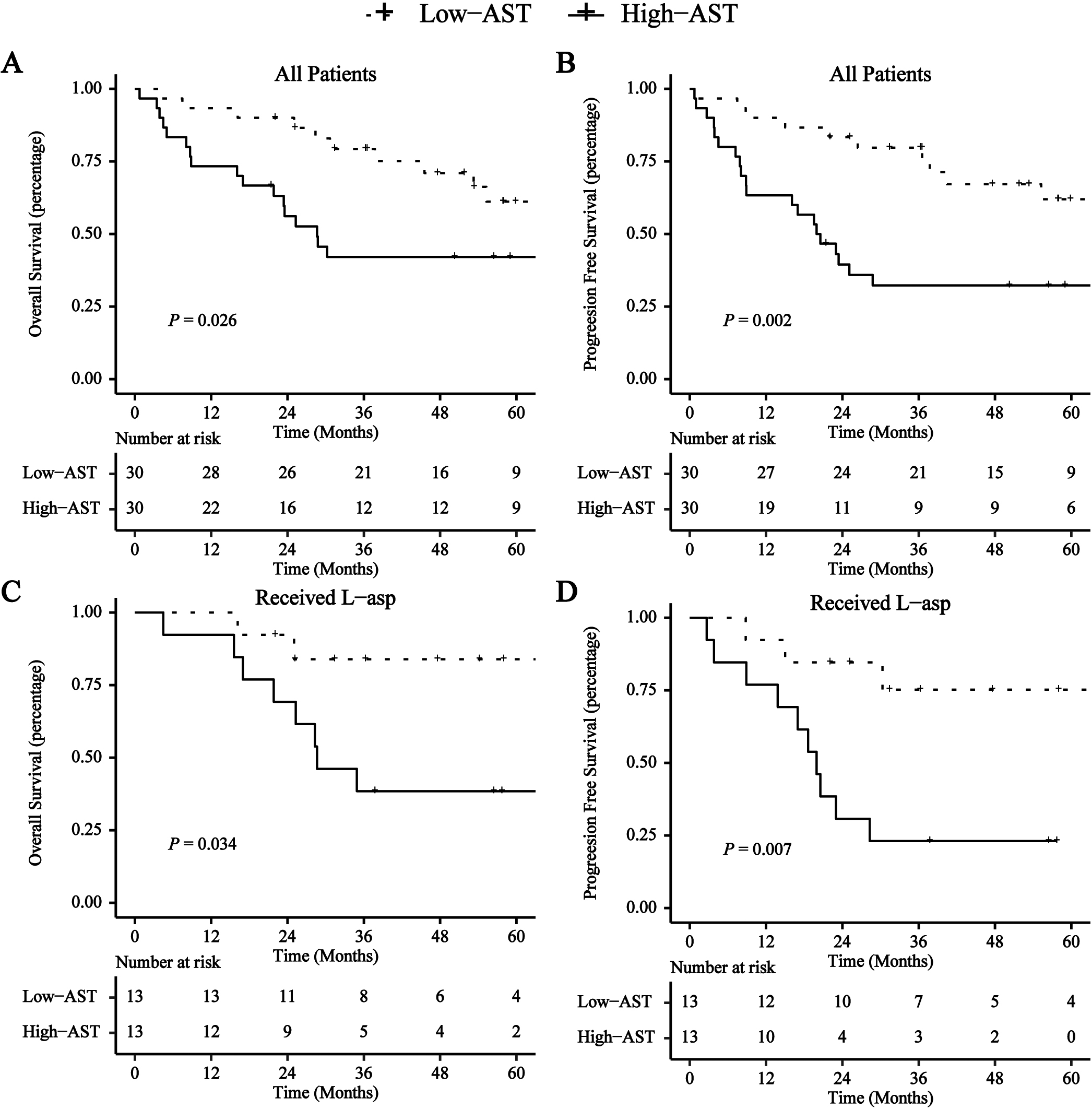
We performed PSM analysis using factors including LDH, ALT/AST, ALT, KPI in the L-Asp-based chemotherapy group. After PSM, the differences for all covariates between the low- (
3.4Subgroup analyses
We performed a subgroup analysis based on patients’ baseline characteristics and treatment, including age, gender, ECOG score, Ann Arbor stage, B symptoms, extranodal involvement site, regional lymph node involvement, subtype, RT, and L-Asp. Figure 3 showed that OS and PFS were shorter in almost all subgroups with a high AST level than those with a low AST. The HRs were 1.575 to 3.916 and 1.803 to 3.730, respectively.
Figure 3.
Forest plot depicting the HRs of AST in different risk subgroups for OS and PFS.
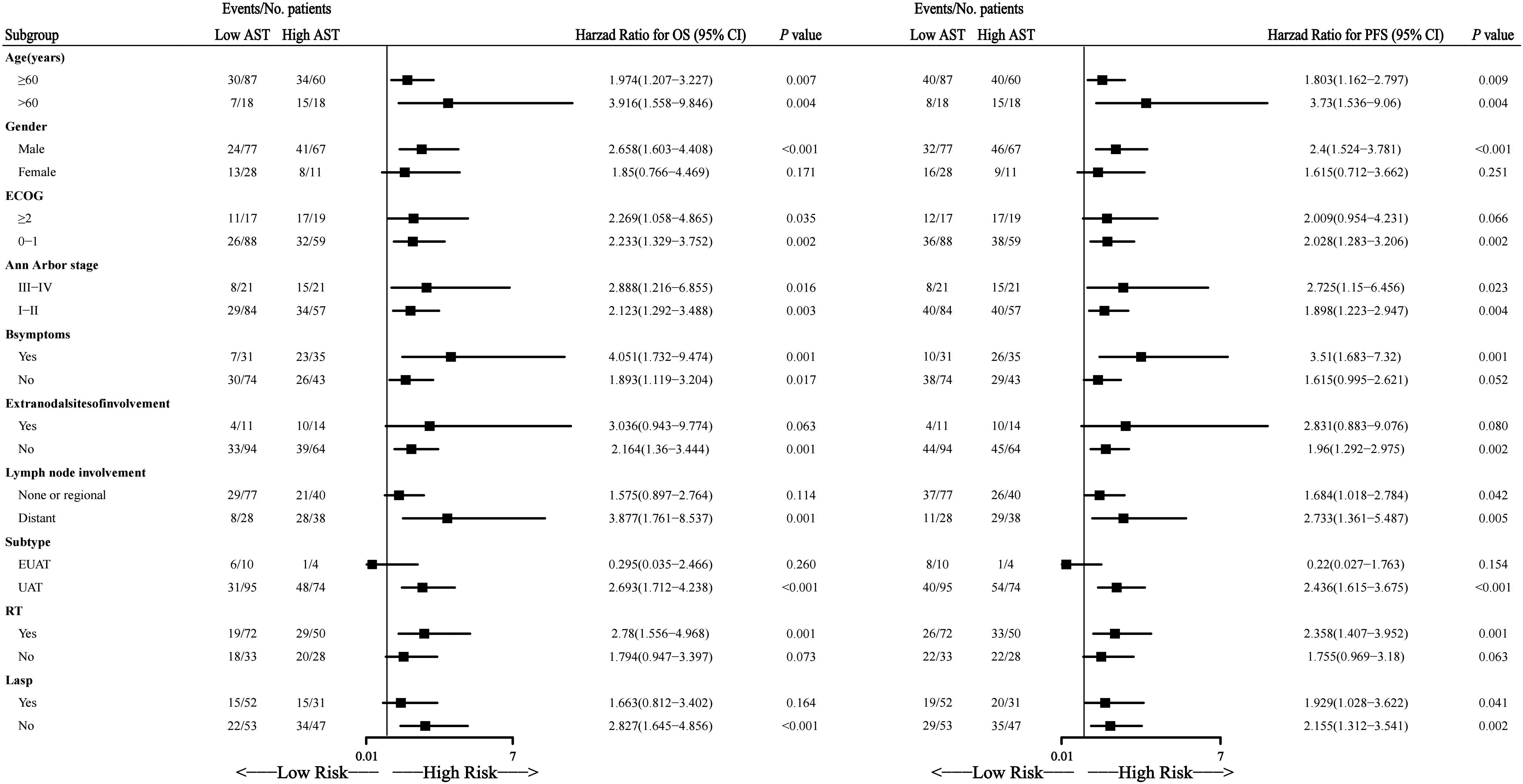
Figure 4.
Time-dependent ROC curve for predicting ENKTL patients’ OS.
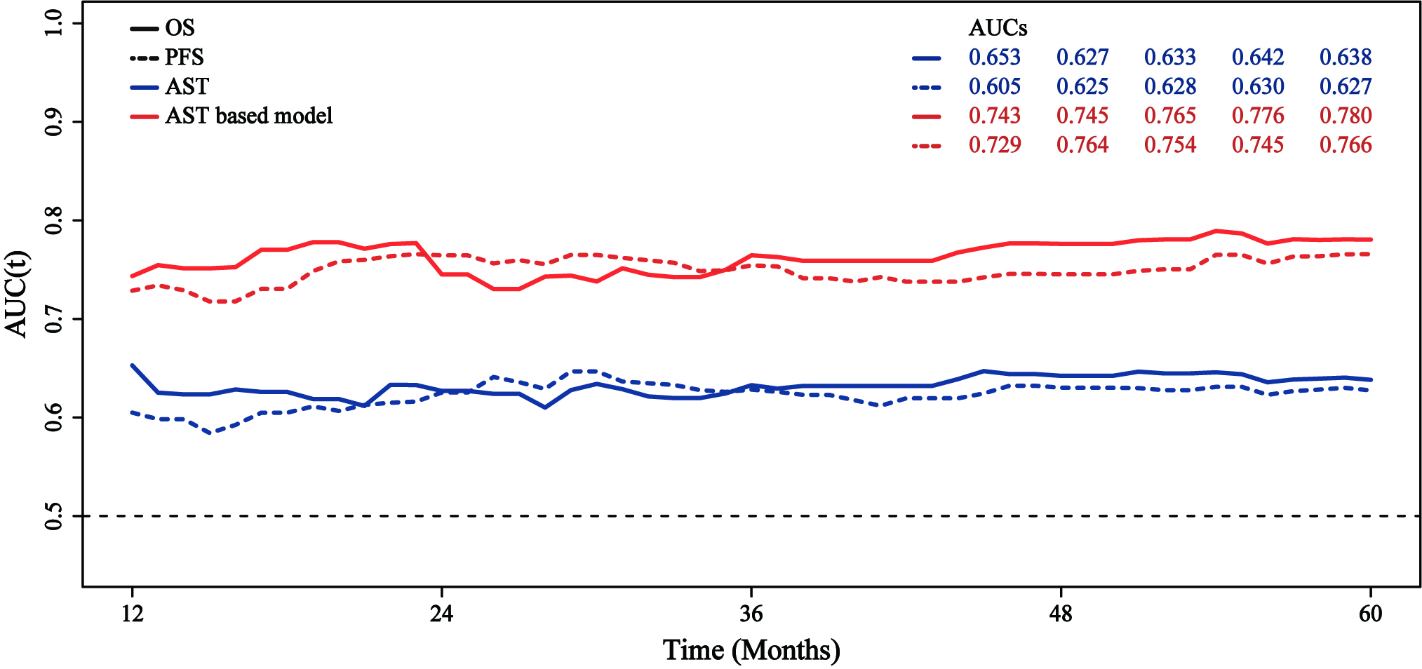
3.5Prognostic value of AST
Accordingly, time-dependent receiver operating characteristics (ROC) curves revealed that AST was a powerful predictor for ENKTL with the area under the curve (AUC) was 0.653, 0.627, 0.633, 0.642, and 0.638 for OS at 12, 24, 36, 48, and 60 months, and 0.605, 0.625, 0.628, 0.630, and 0.627 for PFS at 12, 24, 36, 48, and 60 months, respectively (Fig. 4).
The time-dependent AUCs calculated with 5-fold cross validation was used to assess the performance of the AST on the training cohort and the validation cohort, respectively. When predicting 12, 24, 36, 48, and 60 months OS, the average AUC in the training cohort was 0.654, 0.627, 0.633, 0.643, and 0.639, respectively, and 0.675, 0.627, 0.637, 0.649, and 0.646 in the validation cohort, respectively. When predicting PFS at 12, 24, 36, 48 and 60 months, the average AUC was 0.605, 0.626, 0.628, 0.631, and 0.628 in the training set, and 0.605, 0.628, 0.631, 0.638, and 0.631 in the validation set, respectively (Supplemental Table 2).
AST-based model for predicting OS (Fig. S1) and PFS (Fig. S2) was built based on the multivariate Cox regression model. The time-dependent ROC curve showed that the AST-based model could more effectively predict the OS and PFS of patients. The time-dependent ROC curves revealed that AST was a powerful predictor for ENKTL with the area under the curve (AUC) was 0.743, 0.745, 0.765, 0.776, and 0.780 for OS at 12, 24, 36, 48, and 60 months, and 0.729, 0.764, 0.754, 0.745, and 0.766 for PFS at 12, 24, 36, 48, and 60 months, respectively (Fig 4).
4.Discussion
In this study, we first presented that the serum AST was a prognostic indicator for patients with ENKTL. Recently, some studies reported that AST was increasingly associated with the outcomes of some malignancies, such as non-small cell lung cancer, multiple myeloma, breast cancer, and pancreatic cancer [19, 22, 30, 31]. Furthermore, AST could be a prognostic indicator integrated with ALT, another important circulating transaminase [32, 33]. The ratio of AST to lymphocyte also plays an essential part in tumor prognosis [18, 34]. A study in non-Hodgkin lymphoma showed that higher AST levels predicted a worse prognosis in DLBCL [23]. ENKTL is also a subtype of non-Hodgkin lymphoma, and we hypothesized that AST may be associated with the prognosis of NKT patients. Our results in ENKTL patients were consistent with this finding.
The results of a large-scale study including 416,122 patients showed that elevated AST was not only significantly associated with death from all causes of non-liver disease (
Many studies conventionally evaluated the predictive values of IPI, KPI, and Ann Arbor scores for ENKTL patients. However, the application values still need to be further discussed. Previous studies have shown that most patients were categorized as the low-risk group based on IPI and KPI score and classified as early-stage based on the Ann Arbor Staging System [36]. The disproportionate distribution failed to achieve precise prediction and appropriate clinical guidance. AST helped identify patients with unfavorable outcomes in the low-risk group categorized by the above score systems.
In addition, our finding indicated that higher AST was associated with unfavorable OS and PFS in patients who received L-Asp-based chemotherapy. The efficacy of conventional CHOP (cyclophosphamide, doxorubicin, vincristine, and prednisone) or CHOP-based chemotherapy was limited because of the resistance even when followed by radiotherapy [37, 38]. A meta-analysis showed that L-Asp-based chemotherapy significantly improved complete response (CR) and overall response rate (ORR) of early-stage and advanced-stage ENKTL patients compared with L-Asp-absent regimen [39] and was reported to have more than 80% response rates in patients with refractory or relapsed ENKTL [40, 41]. In this study, we also showed that AST was an independent prognostic factor in receiving L-Asp-based chemotherapy patients.
This study has the following limitations. Due to the .retrospective analysis of a limited number of patients, we need to determine the diagnostic value and further validation through large-scale prospective studies. Besides, further investigations are required to delineate the mechanisms.
5.Conclusion
AST could be an influential prognostic factor for patients with ENKTL.
Funding
This research was funded by the Applied Basic Research Projects of Shanxi Province [No. 20210302124 598], and the Fund Program for the Scientific Activities of Selected Returned Overseas Professionals in Shanxi Province (Department of Resource and Social Security of Shanxi Province No. [2019]1176), the Research Project Supported by Shanxi Scholarship Council of China No. [2022]210, the Key Research and Development (R&D) Projects of Shanxi Province [No. 201803D421054], Wu Jieping Medical Foundation No. 320.6750.2022-1-53, Lianyungang Yixing Medical Health Foundation, and the Four “Batches” Innovation Project of Invigorating Medical through Science and Technology of Shanxi Province No. [2022]37.
Author contributions
Conception: Cao J, Sun R.
Interpretation or analysis of data: Yao NN, Hou Q, Liang Y, Cao X, Sun B, Wei L.
Preparation of the manuscript: Yao NN, Hou Q, Liang Yu.
Revision for important intellectual content: Sun R, Cao J.
Data availability statement
The data of this study are available from the corresponding author upon reasonable request.
Supplementary data
The supplementary files are available to download from http://dx.doi.org/10.3233/CBM-230068.
Conflict of interest
All authors declare no competing interests.
References
[1] | S.H. Swerdlow, E. Campo, S.A. Pileri, N.L. Harris, H. Stein, R. Siebert, R. Advani, M. Ghielmini, G.A. Salles, A.D. Zelenetz and E.S. Jaffe, The 2016 revision of the World Health Organization classification of lymphoid neoplasms, Blood 127: ((2016) ), 2375–90. |
[2] | B.M. William and J.O. Armitage, International analysis of the frequency and outcomes of NK/T-cell lymphomas, Best Pract Res Clin Haematol 26: ((2013) ), 23–32. |
[3] | D.H. Yoon, S.J. Kim, S.H. Jeong, D.Y. Shin, S.H. Bae, J. Hong, S.K. Park, H.Y. Yhim, D.H. Yang, H. Lee, H.J. Kang, M.H. Lee, H.S. Eom, J.Y. Kwak, J.H. Lee, C. Suh and W.S. Kim, Phase II trial of concurrent chemoradiotherapy with L-asparaginase and MIDLE chemotherapy for newly diagnosed stage I/II extranodal NK/T-cell lymphoma, nasal type (CISL-1008), Oncotarget 7: ((2016) ), 85584–85591. |
[4] | B. Cahan and Y.J. Chen, Modern Radiation Therapy for Extranodal Lymphomas: Field and Dose Guidelines From the International Lymphoma Radiation Oncology Group: In Regard to Yahalom et al, Int J Radiat Oncol Biol Phys 93: ((2015) ), 471–2. |
[5] | J. Lee, C. Suh, Y.H. Park, Y.H. Ko, S.M. Bang, J.H. Lee, D.H. Lee, J. Huh, S.Y. Oh and H.-C. Kwon, Extranodal natural killer T-cell lymphoma, nasal-type: a prognostic model from a retrospective multicenter study, Journal of Clinical Oncology 24: ((2006) ), 612–618. |
[6] | C.S. Chim, S.Y. Ma, W.Y. Au, C. Choy, A.K. Lie, R. Liang, C.C. Yau and Y.L. Kwong, Primary nasal natural killer cell lymphoma: long-term treatment outcome and relationship with the International Prognostic Index, Blood 103: ((2004) ), 216–21. |
[7] | W.Y. Au, D.D. Weisenburger, T. Intragumtornchai, S. Nakamura, W.S. Kim, I. Sng, J. Vose, J.O. Armitage and R. Liang, Clinical differences between nasal and extranasal natural killer/T-cell lymphoma: a study of 136 cases from the International Peripheral T-Cell Lymphoma Project, Blood 113: ((2009) ), 3931–7. |
[8] | Y. Yang, J.Z. Cao, S.M. Lan, J.X. Wu, T. Wu, S.Y. Zhu, L.T. Qian, X.R. Hou, F.Q. Zhang, Y.J. Zhang, Y. Zhu, L.M. Xu, Z.Y. Yuan, S.N. Qi and Y.X. Li, Association of Improved Locoregional Control With Prolonged Survival in Early-Stage Extranodal Nasal-Type Natural Killer/T-Cell Lymphoma, JAMA Oncol 3: ((2017) ), 83–91. |
[9] | J. Huang, W. Jiang, T. Lin, Y. Huang, R. Xu, H. Huang and Z. Li, Absolute lymphocyte count is a novel prognostic indicator in extranodal natural killer/T-cell lymphoma, nasal type, Annals of Oncology 22: ((2011) ), 149–155. |
[10] | J. Cao, S. Lan, L. Shen, H. Si, H. Xiao, Q. Yuan, X. Li, H. Li and R. Guo, Hemoglobin level, a prognostic factor for nasal extranodal natural killer/T-cell lymphoma patients from stage I to IV: A validated prognostic nomogram, Sci Rep 7: ((2017) ), 10982. |
[11] | Y. Yang, Y. Zhang, Y. Zhu, J. Cao, Z. Yuan, L. Xu, J. Wu, W. Wang, T. Wu and B. Lu, Prognostic nomogram for overall survival in previously untreated patients with extranodal NK/T-cell lymphoma, nasal-type: a multicenter study, Leukemia 29: ((2015) ), 1571–1577. |
[12] | Y. Yang, Y. Zhu, J.-Z. Cao, Y.-J. Zhang, L.-M. Xu, Z.-Y. Yuan, J.-X. Wu, W. Wang, T. Wu and B. Lu, Risk-adapted therapy for early-stage extranodal nasal-type NK/T-cell lymphoma: analysis from a multicenter study, Blood, The Journal of the American Society of Hematology 126: ((2015) ), 1424–1432. |
[13] | S.J. Kim, D.H. Yoon, A. Jaccard, W.J. Chng, S.T. Lim, H. Hong, Y. Park, K.M. Chang, Y. Maeda and F. Ishida, A prognostic index for natural killer cell lymphoma after non-anthracycline-based treatment: a multicentre, retrospective analysis, The Lancet Oncology 17: ((2016) ), 389–400. |
[14] | K. Xie, C.H. Chen, S.P. Tsai, P.J. Lu, H. Wu, Y. Zeng, Y. Ye, H. Tu, C. Wen, M. Huang, Y. Zhang, J.H. Lee, M.K. Tsai, C.P. Wen and X. Wu, Loss of Life Expectancy by 10 Years or More From Elevated Aspartate Aminotransferase: Finding Aspartate Aminotransferase a Better Mortality Predictor for All-Cause and Liver-Related than Alanine Aminotransferase, Am J Gastroenterol 114: ((2019) ), 1478–1487. |
[15] | V. Arndt, H. Brenner, D. Rothenbacher, B. Zschenderlein, E. Fraisse and T.M. Fliedner, Elevated liver enzyme activity in construction workers: prevalence and impact on early retirement and all-cause mortality, Int Arch Occup Environ Health 71: ((1998) ), 405–12. |
[16] | R. Scherzer, S.B. Heymsfield, D. Rimland, W.G. Powderly, P.C. Tien, P. Bacchetti, M.G. Shlipak and C. Grunfeld, Association of serum albumin and aspartate transaminase with 5-year all-cause mortality in HIV/hepatitis C virus coinfection and HIV monoinfection, Aids 31: ((2017) ), 71–79. |
[17] | T.H. Lee, W.R. Kim, J.T. Benson, T.M. Therneau and L.J. Melton, 3rd, Serum aminotransferase activity and mortality risk in a United States community, Hepatology 47: ((2008) ), 880–7. |
[18] | J. Jin, P. Zhu, Y. Liao, J. Li, W. Liao and S. He, Elevated preoperative aspartate aminotransferase to lymphocyte ratio index as an independent prognostic factor for patients with hepatocellular carcinoma after hepatic resection, Oncotarget 6: ((2015) ), 19217–27. |
[19] | S.L. Chen, N. Xue, M.T. Wu, H. Chen, X. He, J.P. Li, W.L. Liu and S.Q. Dai, Influence of Preoperative Serum Aspartate Aminotransferase (AST) Level on the Prognosis of Patients with Non-Small Cell Lung Cancer, Int J Mol Sci 17: ((2016) ). |
[20] | J.M. Thornburg, K.K. Nelson, B.F. Clem, A.N. Lane, S. Arumugam, A. Simmons, J.W. Eaton, S. Telang and J. Chesney, Targeting aspartate aminotransferase in breast cancer, Breast Cancer Res 10: ((2008) ), R84. |
[21] | C.D. Witjes, I.J. JN, A.A. van der Eijk, B.E. Hansen, C. Verhoef and R.A. de Man, Quantitative HBV DNA and AST are strong predictors for survival after HCC detection in chronic HBV patients, Neth J Med 69: ((2011) ), 508–13. |
[22] | T. Kiba, T. Ito, T. Nakashima, Y. Okikawa, M. Kido, A. Kimura, K. Kameda, F. Miyamae, S. Tanaka, M. Atsumi, Y. Sumitani, Y. Shitakubo and H. Niimi, Bortezomib and dexamethasone for multiple myeloma: higher AST and LDH levels associated with a worse prognosis on overall survival, BMC Cancer 14: ((2014) ), 462. |
[23] | T.-X. Lu, S. Wu, D.-Y. Cai, T.-T. Hong, Y. Zhang, H.-Q. Gao, H.-Y. Hua and X.-H. Wu, Prognostic significance of serum aspartic transaminase in diffuse large B-cell lymphoma, BMC Cancer 19: ((2019) ), 1–9. |
[24] | T.M. Kim and D.S. Heo, Extranodal NK / T-cell lymphoma, nasal type: new staging system and treatment strategies, Cancer Sci 100: ((2009) ), 2242–8. |
[25] | M. Shipp, A predictive model for aggressive non-Hodgkin’s lymphoma. The international non-Hodgkin’s lymphoma prognostic factors project, N Engl j Med 329: ((1993) ), 987–994. |
[26] | J. Lee, C. Suh, Y.H. Park, Y.H. Ko, S.M. Bang, J.H. Lee, D.H. Lee, J. Huh, S.Y. Oh, H.C. Kwon, H.J. Kim, S.I. Lee, J.H. Kim, J. Park, S.J. Oh, K. Kim, C. Jung, K. Park and W.S. Kim, Extranodal natural killer T-cell lymphoma, nasal-type: a prognostic model from a retrospective multicenter study, J Clin Oncol 24: ((2006) ), 612–8. |
[27] | A. Gallamini, C. Stelitano, R. Calvi, M. Bellei, D. Mattei, U. Vitolo, F. Morabito, M. Martelli, E. Brusamolino and E. Iannitto, Peripheral T-cell lymphoma unspecified (PTCL-U): a new prognostic model from a retrospective multicentric clinical study, Blood 103: ((2004) ), 2474–2479. |
[28] | C. Contal and J. O’Quigley, An application of changepoint methods in studying the effect of age on survival in breast cancer, Computational statistics & data analysis 30: ((1999) ), 253–270. |
[29] | N. Simon, J. Friedman, T. Hastie and R. Tibshirani, Regularization Paths for Cox’s Proportional Hazards Model via Coordinate Descent, J Stat Softw 39: ((2011) ), 1–13. |
[30] | A. Birtle, P. Lawton, M. Chaundary, P. Richman and B. McFarlane, Unexplained high AST in locally advanced breast cancer, Breast J 9: ((2003) ), 505–6. |
[31] | D.D. Stocken, A.B. Hassan, D.G. Altman, L.J. Billingham, S.R. Bramhall, P.J. Johnson and N. Freemantle, Modelling prognostic factors in advanced pancreatic cancer, Br J Cancer 99: ((2008) ), 883–93. |
[32] | H. Huang, X.P. Wang, X.H. Li, H. Chen, X. Zheng, J.H. Lin, T. Kang, L. Zhang and P.S. Chen, Prognostic value of pretreatment serum alanine aminotransferase/aspartate aminotransferase (ALT/AST) ratio and gamma glutamyltransferase (GGT) in patients with esophageal squamous cell carcinoma, BMC Cancer 17: ((2017) ), 544. |
[33] | A. Bezan, E. Mrsic, D. Krieger, T. Stojakovic, K. Pummer, R. Zigeuner, G.C. Hutterer and M. Pichler, The Preoperative AST/ALT (De Ritis) Ratio Represents a Poor Prognostic Factor in a Cohort of Patients with Nonmetastatic Renal Cell Carcinoma, J Urol 194: ((2015) ), 30–5. |
[34] | L.-Y. Zhao, D.-D. Yang, X.-K. Ma, M.-M. Liu, D.-H. Wu, X.-P. Zhang, D.-Y. Ruan, J.-X. Lin, J.-Y. Wen and J. Chen, The prognostic value of aspartate aminotransferase to lymphocyte ratio and systemic immune-inflammation index for overall survival of hepatocellular carcinoma patients treated with palliative treatments, Journal of Cancer 10: ((2019) ), 2299. |
[35] | S.E. Elf and J. Chen, Targeting glucose metabolism in patients with cancer, Cancer 120: ((2014) ), 774–80. |
[36] | H. Hong, Y. Li, S.T. Lim, C. Liang, H. Huang, P. Yi, T. Wu, X. Du, M. Zhang and J. Wang, A proposal for a new staging system for extranodal natural killer T-cell lymphoma: a multicenter study from China and Asia Lymphoma Study Group, Leukemia 34: ((2020) ), 2243–2248. |
[37] | S.J. Kim and W.S. Kim, Treatment of localized extranodal NK/T cell lymphoma, nasal type, Int J Hematol 92: ((2010) ), 690–6. |
[38] | W.S. Kim, S.Y. Song, Y.C. Ahn, Y.H. Ko, C.H. Baek, D.Y. Kim, S.S. Yoon, H.G. Lee, W.K. Kang, H.J. Lee, C.H. Park and K. Park, CHOP followed by involved field radiation: is it optimal for localized nasal natural killer/T-cell lymphoma? Ann Oncol 12: ((2001) ), 349–52. |
[39] | V.S. Pokrovsky and D. Vinnikov, L-Asparaginase for newly diagnosed extra-nodal NK/T-cell lymphoma: systematic review and meta-analysis, Expert Review of Anticancer Therapy 17: ((2017) ), 759–768. |
[40] | W. Yong, W. Zheng, J. Zhu, Y. Zhang, X. Wang, Y. Xie, N. Lin, B. Xu, A. Lu and J. Li, L-asparaginase in the treatment of refractory and relapsed extranodal NK/T-cell lymphoma, nasal type, Annals of hematology 88: ((2009) ), 647–652. |
[41] | A. Jaccard, N. Gachard, P. Coppo, F. Morschhauser, L. Galicier, L. Ysebaert, P. Lenain, B. Corront, F. Suarez and J. Feuillard, A prospective phase II trial of an L-asparaginase containing regimen in patients with refractory or relapsing extra nodal NK/T-cell lymphoma, American Society of Hematology, (2008) . |


