
Evaluation of the Effects of Opium on the Expression of SOX2 and OCT4 in Wistar Rat Bladder
Abstract
BACKGROUND:
Bladder cancer is a malignancy greatly affected by behavioral habits. The aim of this study was to examine the effect of opium on changes in the expression of OCT4 and SOX2 in the bladder tissue of rats.
METHOD:
Thirty six rats were divided into six groups: 24 rats in the addicted group received morphine and opium for 4 months with 12 rats in the control group. Blood testing was done for the evaluation of CBC, MDA, and TAC. The bladder tissue was removed and checked by histopathological examination. All total RNA was extracted, then cDNAs were synthesized and the OCT4 and SOX2 gene expressions were evaluated by Real-time PCR.
RESULTS:
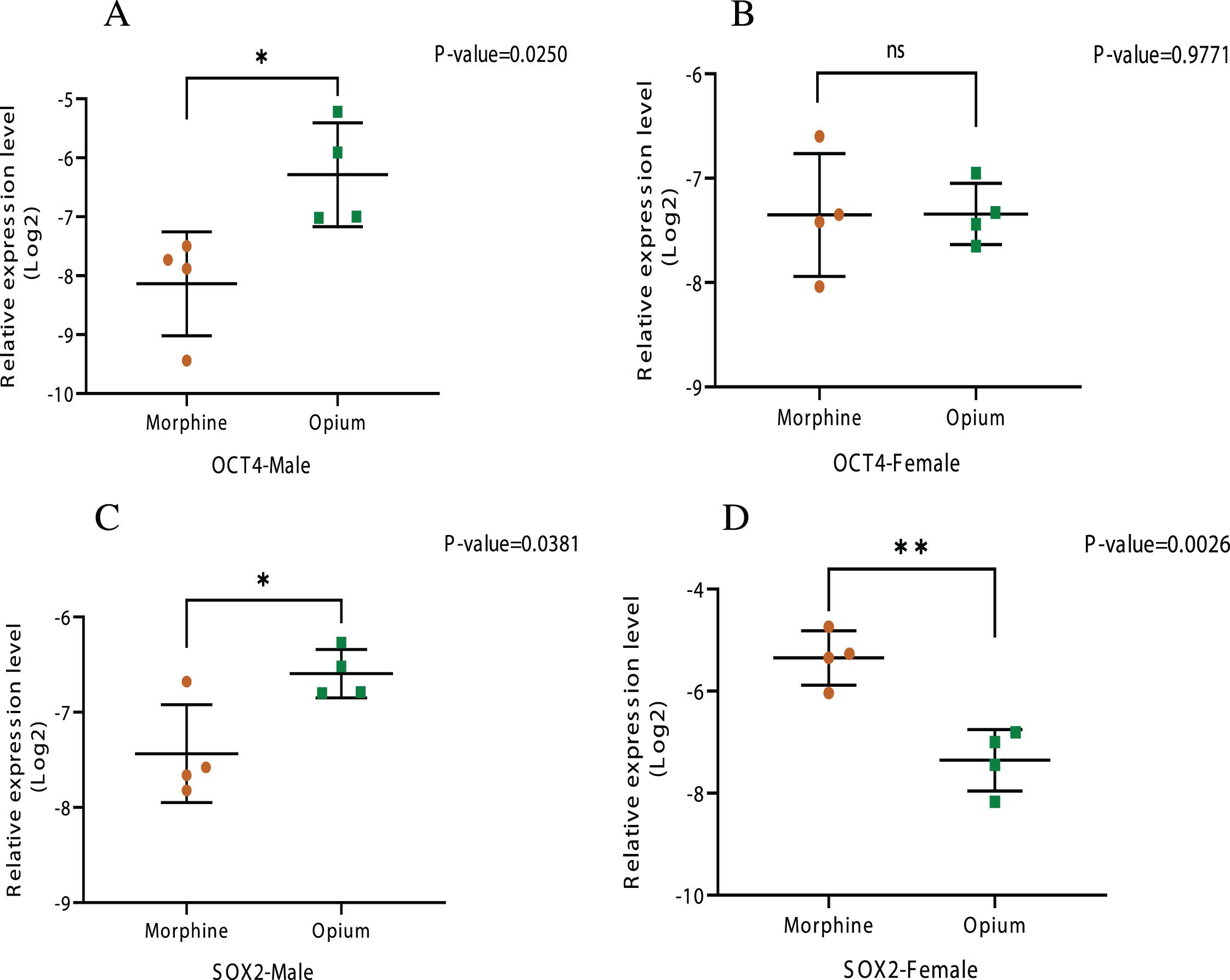
The OCT4 mRNA expression level in the opium group of rats was significantly increased compared to the control group (13.5 and 6.8 fold in males and females respectively). Also, in the morphine group, similar augmentation was detected (3.8 and 6.7 fold in males and females respectively). The SOX2 mRNA over-expression level was seen in the morphine group of both genders as compared to the control group (3.7 and 4.2 fold in male and female respectively) but in the opium group, enhancement of mRNA level was seen only in males (6.6 fold). Opium increases both OCT4 and SOX2 expression more than morphine in male rats, but in female rats, SOX2 is increased more by morphine.
CONCLUSION:
Over expression of OCT4 and SOX2 was observed in rats treated with opium and morphine. Increased OCT4 and SOX2 expression was seen in opium-treated male rats, but in female rats, SOX2 was increased more by morphine.
Abbreviations
BAX Bcl-2-associated X protein
BCl2 B-cell lymphoma 2
CBC complete blood count
cDNAs complementary deoxyribonucleic acid
H & E hematoxylin and eosin
HCT hematocrit
Hgb hemoglobin
MCHC mean corpuscular hemoglobin concentration
MDA malondialdehyde
OCT4 octamer-binding transcription factor 4
PLT platelets
RBC red blood cell
RNA ribonucleic acid
SOX2 SRY-Box Transcription Factor 2
TAC total antioxidant capacity
WBC white blood count
INTRODUCTION
Bladder cancer is the most prevalent genitourinary-system malignancy, and plays a primary role in more than 7% of neoplasms, especially among men [1]. Yearly reports show a further 12 million cases of bladder cancer, putting it in the eleven top-ranked cancers, by incidence, in the world [2] It is the third most common cancer in Iranian men [3] and it occupies the ninth place in prevalence among Iranian women [4]. The risk factors which can lead to bladder cancer include high body mass index [5], genetic factors [6], occupational hazards [7, 8], environment and nutrition [9], and behavioral factors [8, 10–12]. In previous studies, behavioral factors such as smoking and opium addiction were counted as the most important risk factors for several cancers, including bladder cancer [13–15]. Several new treatment strategies are suggested for bladder cancer [16–19]. Meta-analysis agrees that opium consumption is potentially responsible for the development of bladder cancer [20]. The prevalence of opioid consumption ranges from 0.1% to 2% globally [21], and a high prevalence of opioid use has been correlated to Iranians for several hundred years [22]. Although the use of opium is prohibited in Iran, it is the most commonly used drug, and up to 14% of the Iranian population are regular users of opium [23]. Opioids such as morphine, heroin, and opium are the most common form of drugs used in Iran. They can have a negative impact on the immune system [24–26], and have been reported to augment the susceptibility of animals to bacterial and viral infections [27]. They also cause increased metastasis and –as this leads to apoptosis –cell death, and can decrease survival in tumor-bearing animals [28]. Research has also shown apoptosis in the brain and liver of opium-addicted rats [29].
Uncontrolled self-renewal is seen as an essential mechanism in carcinogenesis [30]. By referring to the cancer stem cell (CSC) hypothesis, a batch of cancer cells with stem-cell-like features can sustain a tumor; these batches should be self-renewing and pluripotent [31–33]. The CSCs have an indistinct proliferation, and can also lead to tumorigenesis [34]. The hypothesis is that the single pathway that manages self-renewal in normal stem cells is also employed to manage CSCs in tumors [35]. It is also believed that a member of the POU family, called Octamer 3/4 (Oct 3/4), is a crucial stem-cell marker and an essential transcription factor through human embryogenesis [36]. Recent reports have contained information regarding the presence of Oct 3/4 among differentiated benign and malignant human cells [37]. Sox2 is responsible for encoding transcription factors with a single HMG DNA-binding domain. Another function of this member of the SOX (SRY-Related HMG box) gene family is controlling neural progenitor cells by prohibiting their ability to differentiate [38]. Sox2 expression is also present in various malignant tissues [39].
This study aims to examine the effect of opium on changes in the expression of OCT4 and SOX2, the main controlling genes of the self-renewal pathway, in the bladder tissue of rats.
MATERIALS AND METHODS
This research was carried out in the Animal Laboratory of the Urology Research Center of Tehran Medical University, Tehran, Iran, between October 2021 and September 2022, and approved by the Ethics Research Committee of Tehran University of Medical Sciences (IR.TUMS.SINAHOSPITAL.REC.1399.026). Ethical principles of working with rats were considered in all phases of the research.
Animal care
In this study, 38 4-week old male and female adult Wistar rats weighing between 200 and 220 g each, were purchased from the Pharmacology College of Tehran University, Iran. Four rats were placed in each Plexiglas cage, and given ad libitum access to food and water. The temperature of the laboratory was set at 22±2°C, relative humidity was 45.00±2.00% and the light/dark cycle was 12/12 h. Before commencing the experiments, the rats were allowed to adapt to the lab’s environment for seven days. Maintenance of all animals was in accordance with institutional guidelines for animal care and use and all the experimental protocols were approved by the National Institutes of Health guidance for the care and use of laboratory animals [40].
Exposure of rats to morphine sulfate and powdered opium
In Iran, opium is generally used in one of three different forms; Teriak (crude opium), Sukhteh (remnants of smoked opium or dross), and Shireh (opium juice, an opium product usually made by boiling Teriak or Sukhteh in water, filtering the mixture several times and then evaporating the filtrate) [41].
Morphine sulfate and opium powder (Teriak) (Temad Manufactory, Tehran, Iran), and Naloxone hydrochloride (Tolid Daru company, Tehran, Iran) were used in this study. The drugs were dissolved in sterile 0.9% saline and injected intraperitoneally (i.p.) at a volume of 1 ml/kg.
Two rats were dissected at the beginning of the study. The 36 remaining rats were divided into six groups: 12 rats in the control group (6 females and 6 males) received a single i.p. injection of saline for four months and were treated in the same conditions as the other groups. Rats in the addicted group (6 males and 6 females in each group of morphine and opium) received an initial dose of morphine (2 mg/kg) and opium (2 mg/kg).The morphine dose was then increased every fifteen days with a 4, 8, 10, 15, 20, 25 & 40 mg/kg dose respectively, and the opium dose every 30 days with 10, 20, 40 mg/kg doses respectively, via i.p. injection (a total of 120 days) [40, 42, 43].
To confirm the addiction of the rats, one month after the first injection of morphine and opium, 2 rats in each group received Naloxone randomly (2.5 mg/kg, to confirm addiction) [40, 44] and were placed in a transparent glass box to observe any withdrawal signs such as diarrhea, genital licking, wet-dog shakes, yawing, and sweeping tail movements.
Investigation of serum parameters
Blood samples (5 ml) were taken from rats exposed to opium and morphine from each group before starting the study and immediately after the completion of the project. Nihon Kohden MEK-1305 Celltac α+ cell counter device (automated hematology and Erythrocyte sedimentation rate)ESR(analyzer was used to conduct a complete blood count (CBC) examination. Malondialdehyde (MDA) content was determined by colorimetric method, using a kit purchased from Eagle Biosciences, Inc., USA, according to the method of Satoh [45]. Total Antioxidant Capacity (TAC) was determined using the ELISA technique, using a kit purchased from Cell Biolabs, Inc, San Diego, USA, according to Campbell et al. [46].
Bladder pathology examination
Bladder tissue was removed from the Wistar rats and placed in 10% neutral-buffered formalin. After bladder tissue fixation, the bladder specimens were sent to the comparative histopathology laboratory in the pathology department of the Tehran Medical University, Urology Research Center. According to the standard method, paraffin blocks were processed after molding, and five-micron microscopic incisions from diverse sections were prepared and stained according to the H & E (combination of two histological stains containing hematoxylin and eosin) standard protocol. Observations were performed by Olympus light microscope with ×100, ×200 and ×400 magnifications.
RNA isolation and real-time PCR
All total Ribonucleic acid (RNA) of Wistar rat Bladder tissues was extracted using a High Pure RNA Isolation Kit (Cat. No. 11 828 665 001) and then synthesizing the complementary Deoxyribonucleic acid (cDNAs) (Cat. #RR014A/B). The relative expression of genes was then evaluated using the 2−ΔΔCT method used for calculating relative expression. The primers used for the Rotor gene real-time PCR cycler of OCT4-F mRNA were as follows: forward primer, CCGTGTGAGGTGGAACCTG and reverse primer: CGGTTACAGAACCACACTCG, SOX2 mRNA were as follows: forward primer, CGAGTAGGACATGCTGTAGG and reverse primer: ACATGAACGGCTGGAGCAAC. The forward primer sequence for GAPDH mRNA quantification as housekeeping gene was GGCAAGTTCAACGGCACAG, the reverse primer sequence was CGCCAGTAGACTCCACGAC. The performance of Triplicate qPCR reactions was applied to the samples one by one. We describe the actual time of qPCR condition as 95°C for 10 min in the role of the pre-denature step, after that 40 PCR cycles at 95°C for 30 s, 60°C for 30 s, and 72°C for 30 s. Melting curve analysis was performed, to measure the specificity of the real-time RT-PCR.
Statistical analysis
All experiments were performed three times and showed similar results. At the end of the study, the data was achieved from the graph pad prism 9. Statistical significance was defined at *P < 0.05, **P < 0.01, and ***P < 0.001. one way, two way ANOVA, and T test were performed to evaluate the significance of differences.
RESULTS
Expression of the OCT4 and SOX2 genes
The different mRNA expression levels of the main controlling genes of the self-renewal pathway (OCT4 and SOX2) before and after the administration of opium and morphine were detected by RT-PCR.
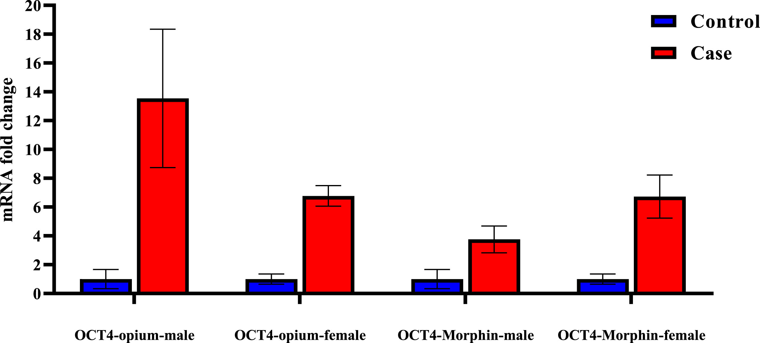
As shown in Fig. 1, the OCT4 mRNA expression level in opium-group rats was significantly increased compared to the control group (13.5 and 6.8 fold in males and females respectively). Similar augmentation was also detected in the morphine group (3.8 and 6.7 fold in males and females respectively).
Fig. 1
OCT4 mRNA fold changes were significantly increased in all groups compared to the control group.
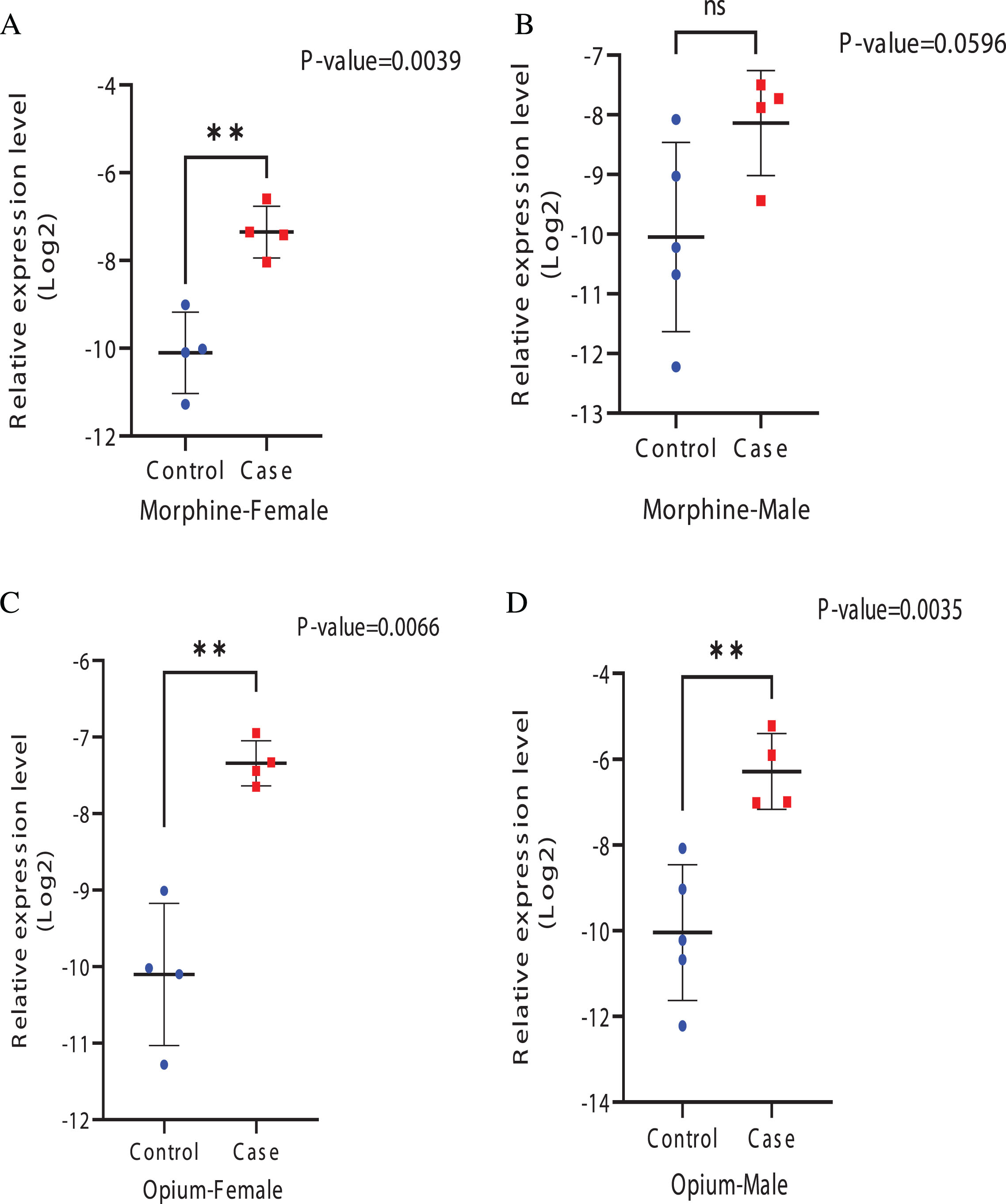
The relative mRNA expression levels of the OCT4 gene were also evaluated by real-time PCR. As shown in Fig. 2, the mRNA expression of OCT4 is detectable for all of the groups studied. Although the mRNA quantities are changing, a comparison of the relative gene-expression levels indicated significant differences among the studied groups (p < 0.05) except in males of the morphine group (p = 0.0596).
Fig. 2
Relative expression levels of OCT4 gene in different groups A) Morphine-Female. B) Morphine-Male. C) Opium-Female. D) Opium-male. There was no significant difference in males of the morphine group, but a noticeable difference was observed in other groups.
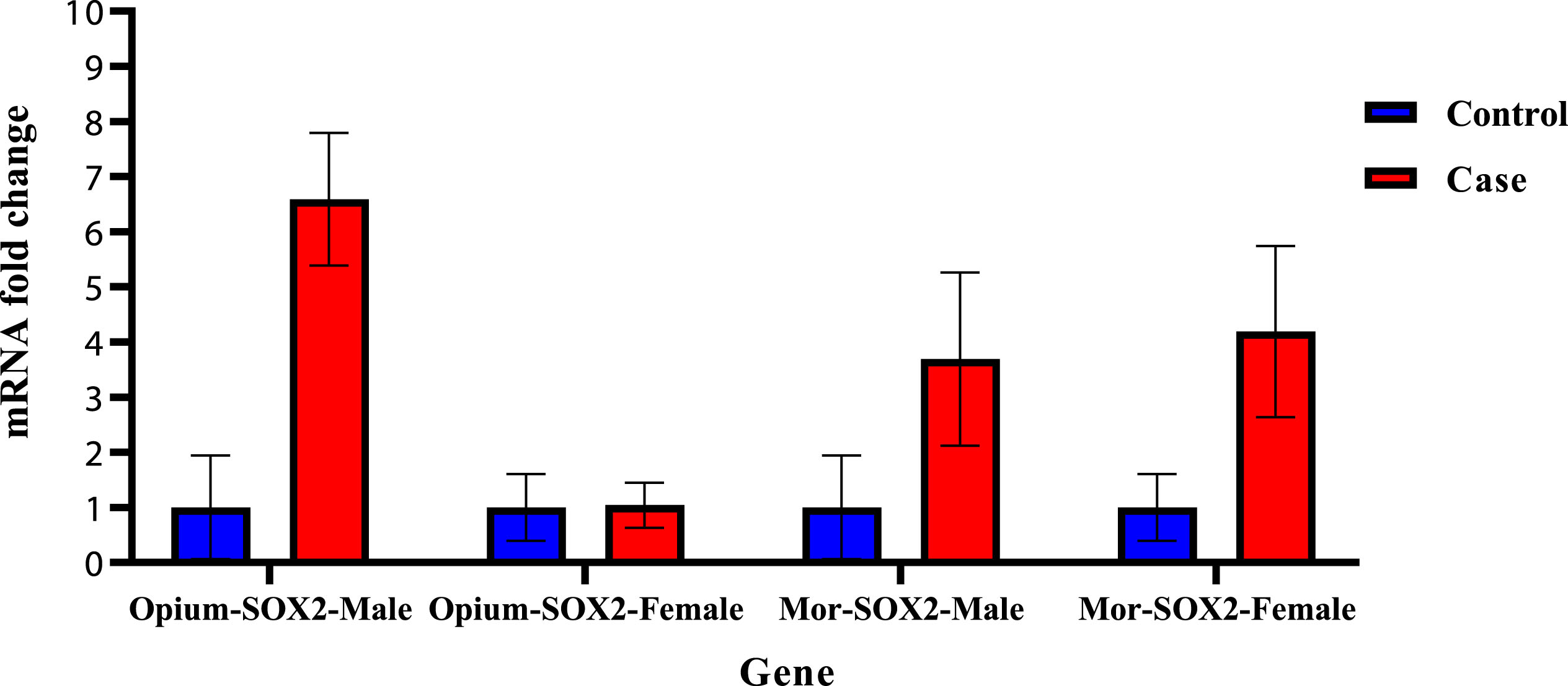
Increase of the SOX2 mRNA expression level was seen in the morphine group of rats of both genders as compared to the control group (3.7 and 4.2 fold in male and female respectively) but in the opium group, enhancement of mRNA level was seen only in males (6.6 fold) (Fig. 3).
Fig. 3
SOX2 mRNA fold change was increased in all groups except females in the opium group.
The relative mRNA expression levels of the SOX2 gene are shown in Fig. 4. As shown in Fig. 4, the mRNA expression of SOX2 is detectable for all of the studied groups. A comparison of the relative gene-expression levels indicated significant differences among the morphine groups. (p < 0.05).
Fig. 4
Relative expression levels of SOX2 gene in case and control groups A) Morphine-Female. B) Morphine-Male. C) Opium-Female. D) Opium-male.
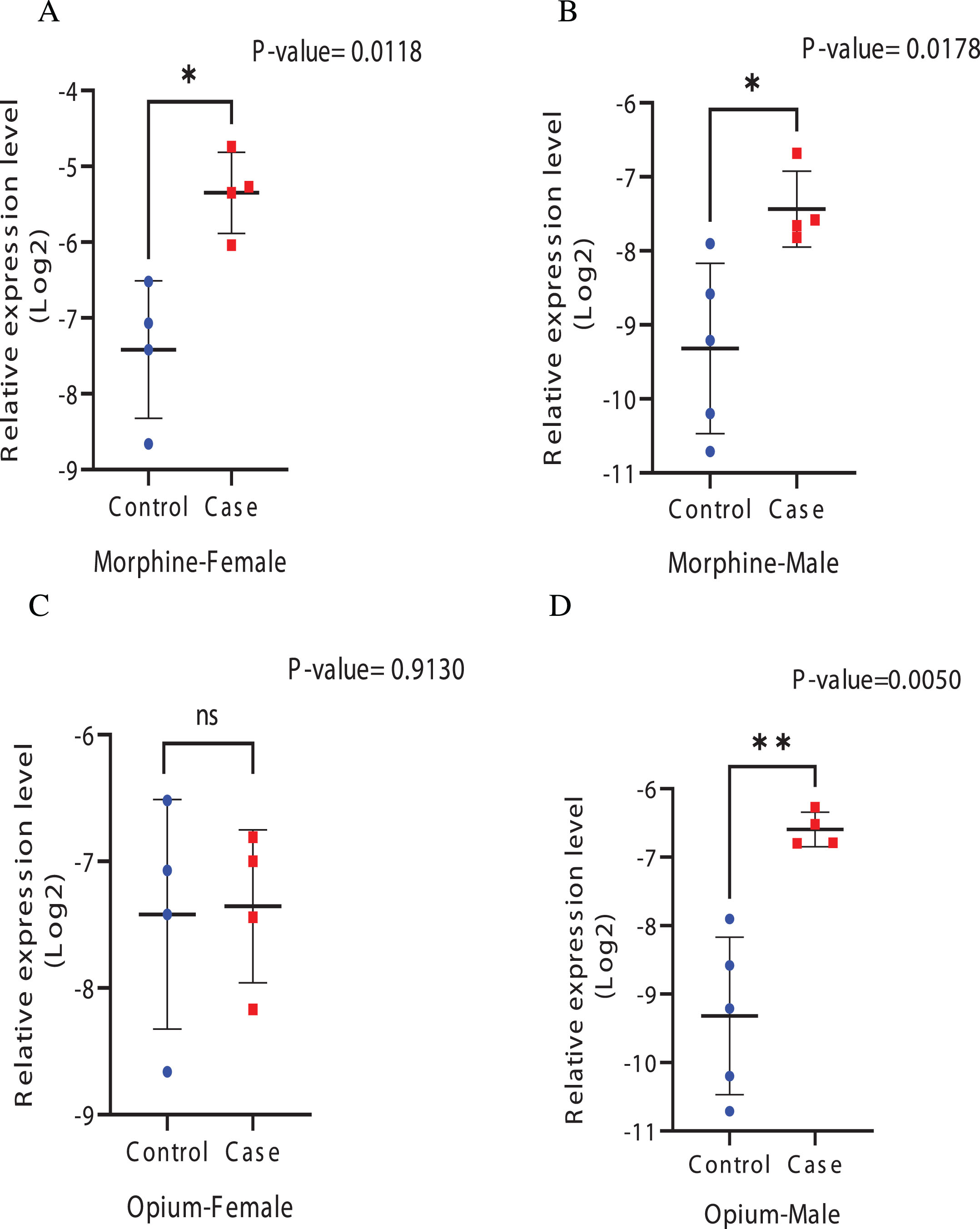
Figure 5 demonstrates that opium increases OCT4 and SOX2 expression more than morphine in male rats but in female rats, SOX2 is increased more by morphine.
Fig. 5
relative expression levels of genes between opium and morphine A) OCT4-Male B) OCT4-Female C) SOX2-Male D) SOX2-Female.
Histopathological findings
In order to confirm our results, various tissues such as the kidney and liver were harvested from the Wistar rats and analyzed with Hematoxylin and Eosin (H&E) staining. We required a fixative in which to soak the tissue sections, and we chose 10% neutral-buffered formalin for this purpose. Tissues were fixed, mounted in paraffin wax, and the slides were prepared before staining with H&E. Staining was carried out on all tissues. A light microscope was then utilized for monitoring the slides in a pathology laboratory, slide observation was also performed using an OPTIKA, C-P2504 microscope.
The results of staining on bladder tissue showed urothelial atrophy in the morphine group (star in Fig. 6) and urothelial cell hyperplasia in the opium group (Arrowhead at Fig. 6). Also, hypertrophy of smooth muscle of the vascular wall and hyperemia were seen in the morphine group as compared with those in the control group (arrows in Fig. 6). Pathology findings for urinary bladder of all rats are also illustrated in Supplementary Table 1.
Fig. 6
Histopathological sections of urinary bladder. Female and male rats, H&E staining. Arrows indicate the urothelial atrophy and star indicates the urothelial cell hyperplasia. Arrows indicate hypertrophy of smooth muscle of vascular wall and hyperemia.
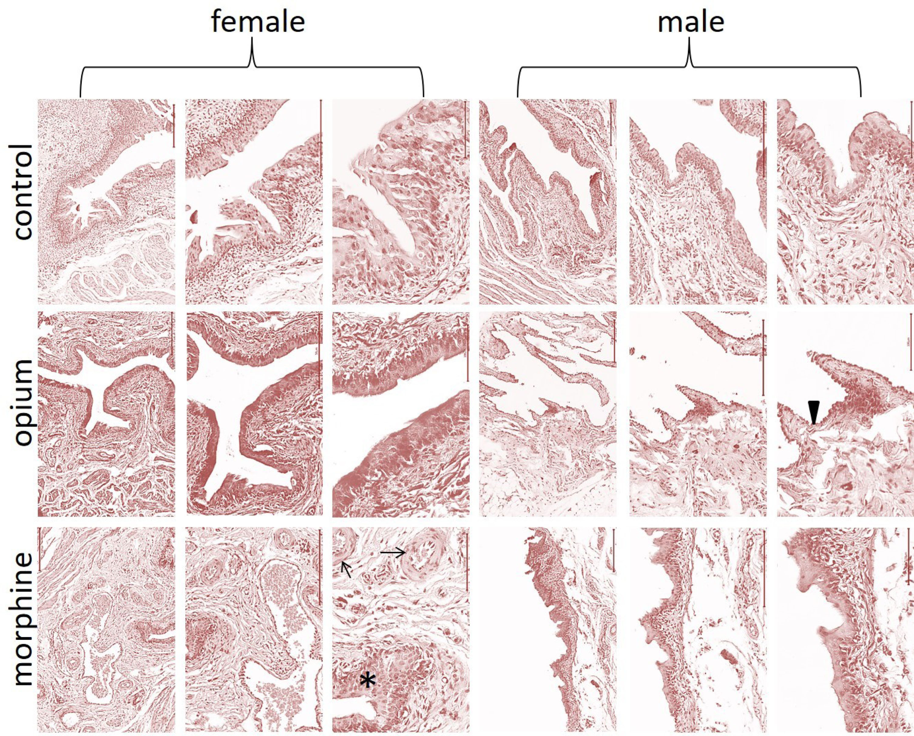
Table 1
Mean values (SD) of CBC, TAC, and MDA of rats from each group
| Variables | Control | Morphine | Opium | p-value | Post-hoc p-values | |||
| CvsM | CvsO | MvsO | ||||||
| WBC | Female | 5.72 (0.42) | 6.86 (1.82) | 5.54 (0.37) | 0.162 | 0.379 | 1 | 0.244 |
| Male | 10.22 (0.80) | 7.00 (1.63) | 7.70 (0.10) | 0.001 | 0.001 | 0.008 | 0.940 | |
| RBC | Female | 7.48 (0.78) | 7.78 (0.80) | 6.78 (0.36) | 0.096 | 1 | 0.386 | 0.114 |
| Male | 8.76 (0.55) | 8.18 (0.40) | 9.60 (0.25) | 0.001 | 0.149 | 0.025 | 0.001 | |
| HGB | Female | 13.86 (0.82) | 14.6 (0.55) | 12.92 (0.44) | 0.004 | 0.253 | 0.102 | 0.003 |
| Male | 14.8 (1.09) | 14.7 (0.47) | 10.10 (0.16) | <0.001 | 1 | <0.001 | <0.001 | |
| HCT | Female | 37.50 (1.64) | 39.80 (0.96) | 34.88 (0.18) | <0.001 | 0.019 | 0.008 | <0.001 |
| Male | 40.88 (0.76) | 39.86 (0.86) | 27.54 (0.42) | <0.001 | 0.124 | <0.001 | <0.001 | |
| MCV | Female | 50.02 (3.08) | 51.02 (1.06) | 51.60 (0.94) | 0.459 | 1 | 0.679 | 1 |
| Male | 46.70 (7.79) | 48.66 (1.72) | 47.62 (1.62) | 0.808 | 1 | 1 | 1 | |
| MCH | Female | 18.48 (0.50) | 17.94 (1.00) | 19.1 (0.89) | 0.128 | 0.969 | 0.779 | 0.141 |
| Male | 16.90 (0.89) | 17.98 (0.71) | 17.6 (0.35) | 0.079 | 0.088 | 0.404 | 1 | |
| MCHC | Female | 36.90 (1.31) | 36.92 (0.72) | 37.00 (1.01) | 0.987 | 1 | 1 | 1 |
| Male | 36.30 (0.94) | 36.94 (0.64) | 45.70 (0.46) | <0.001 | 0.537 | <0.001 | <0.001 | |
| PLT | Female | 692.00 (31.91) | 715.50 (58.23) | 737.00 (61.03) | 0.42 | 1 | 0.591 | 1 |
| Male | 920.00 (18.50) | 715.50 (35.42) | 510.56 (0.53) | <0.001 | <0.001 | <0.001 | <0.001 | |
| RDW | Female | 10.50 (0.87) | 11.60 (0.89) | 11.02 (0.04) | 0.092 | 0.097 | 0.826 | 0.679 |
| Male | 12.20 (0.45) | 12.84 (1.32) | 11.96 (0.21) | 0.250 | 0.714 | 1 | 0.340 | |
| TAC | Female | 0.98 (0.28) | 2.06 (0.68) | 2.09 (0.49) | 0.007 | 0.018 | 0.015 | 1 |
| Male | 1.20 (0.45) | 2.06 (0.58) | 2.40 (0.23) | 0.003 | 0.029 | 0.003 | 0.746 | |
| MDA | Female | 8.54 (0.54) | 9.14 (0.48) | 5.96 (0.37) | <0.001 | 0.195 | <0.001 | <0.001 |
| Male | 6.06 (0.13) | 9.34 (0.29) | 8.21 (0.40) | <0.001 | <0.001 | <0.001 | <0.001 | |
p-values from ANOVA, CvsM = control versus morphine; CvsO = control versus opium; MvsO = morphine versus opium.
Serological findings
The mean values of the serological parameters for rats from each of the six groups were evaluated (Table 1). Values included are: CBC, total antioxidant capacity (TAC), and Malondialdehyde (MDA). A comparison was made of these parameters in the various groups (Fig. 7).
Fig. 7
Comparison of serological parameters in various groups; mean (95% CI).
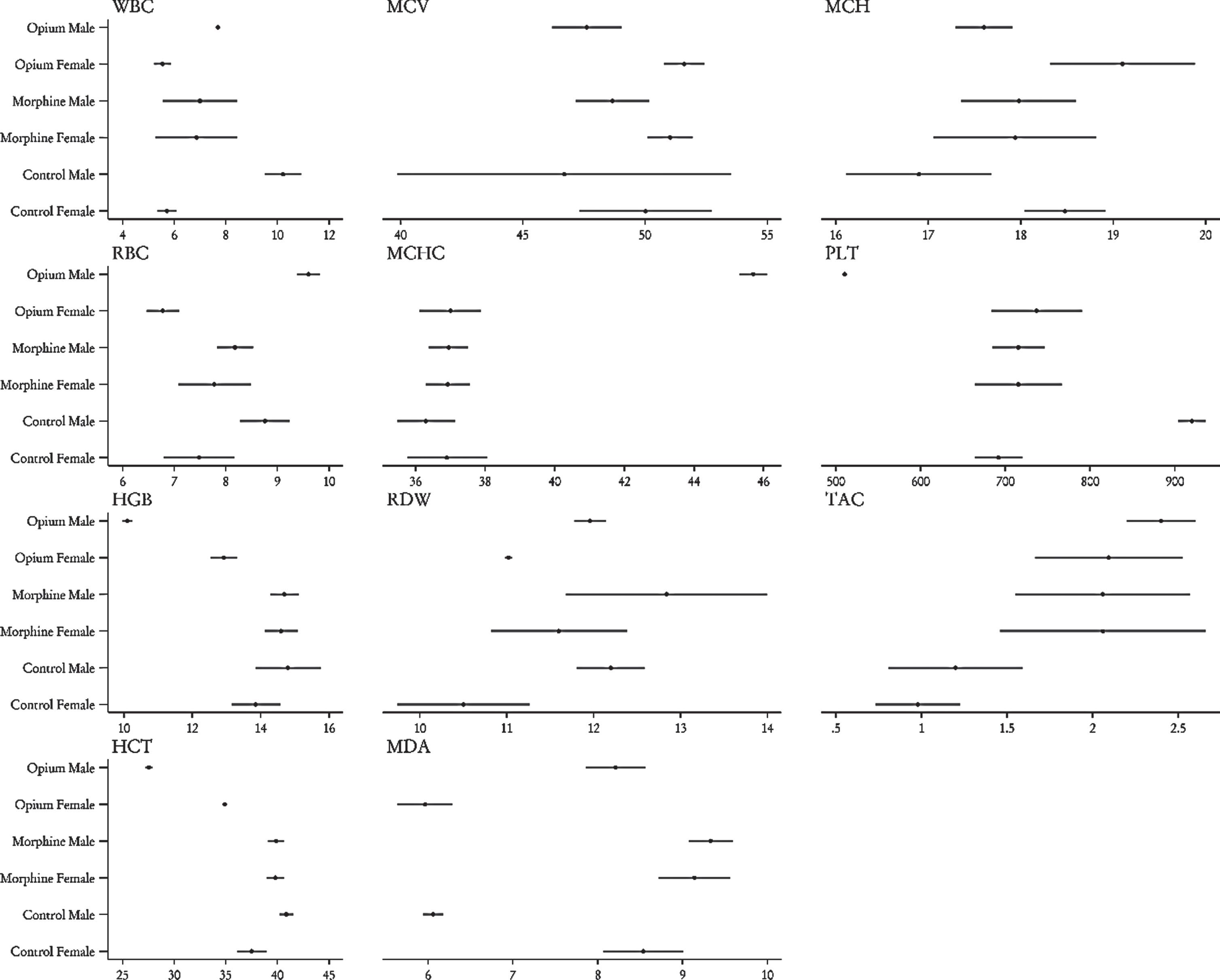
As regards male rats, observations indicated that opium treatment led to a reduction in white blood cells (WBC), hemoglobin (Hgb), hematocrit (HCT), and platelets (PLT), Increases in red blood cells (RBC), mean cell hemoglobin concentration (MCHC), MDA, and TAC were observed as compared to the control group. However, opium treatment caused a decrease in HCT and MDA levels and an increase in TAC levels in female rats as compared to the control group.
By comparing opium and morphine in detail, it was established that opium raises RBC and MCHC and causes a decline in Hgb, HCT, PLT, and MDA more than morphine in male rats, but that in female rats, only Hgb, HCT, and MDA are decreased more by opium.
DISCUSSION
Bladder cancer is the most prevalent malignancy of the genitourinary system [1]. Behavioral factors such as smoking and opium addiction are counted as the most important risk factors for several cancers, including bladder cancer [13, 14]. The prevalence of opioid consumption ranges from 0.1% to 2% of population globally [21]. In the Middle East and Asia, illicit opioid use exists across a spectrum between heroin and opium. Among 204 opioid-use disorder patients, in 56 (29.3%) opium was the primary substance of choice [47]. Based on a national survey conducted among Iranian workers, opium proved to be the most popular substance, and 2.7% of workers were using it [48], and it is estimated that about 20% of the adult population of Iran consumes it [49]. In recent years, there have also been reports of the presence of OCT4 and SOX2 expression in a number of malignant tissues. In this study, we found that morphine and opium significantly increased OCT4 expression in both male and female rats. SOX2 was expressed more in morphine cases as compared to the control group, but in the opium group, this increase was only observed in male rats. In male rats, opium increases OCT4 and SOX2 expression more than morphine. In female rats however, morphine increases SOX2 more than opium.
There are other studies that confirm our results. An increase observed in the mRNA levels of OCT4, SOX2, and Nanog in both MCF-7 and BT549 cells was caused by morphine, as discussed by Niu et al.. They reported that the mRNA levels of OCT4, SOX2, and Nanog were enhanced as compared to untreated controls by:13.08, 10.57, and 19.18 fold in MCF-7 cells, while the initial values reported were: 6.15, 10.37, and 14.92 fold in BT549 cells. We observed a similar effect; the growth in OCT4 expression was reported as 3.8 and 6.7 fold in males and females, and SOX2 expression was increased by 3.7 and 4.2 fold in both males and females in our study. Increases in OCT4 and SOX2 expression were greater in the Niu study as compared to our study. They also confirmed their findings by western blot, showing that the increase in protein levels of OCT4, SOX2, and Nanog in MCF-7 and BT549 cells is dependent on morphine dose, so they recommend that tuning OCT4, SOX2, and Nanog by means of morphine may promote cancer stem cell properties [50].
SOX2, OCT4, and Nanog are stem-cell markers that develop cancer and increase drug resistance [51]. It is believed that stem-cell markers are significantly increased by morphine, so reports agree that the enrichment of stem-cell properties and drug resistance may be caused by long-time morphine use [50].
Gonzalez et al. also agree with these results. In their report, it was observed that SOX2, OCT 4, and Nanog were highly expressed in zebra-fish embryos treated with morphine, as qPCR indicated [52].
SOX2 and OCT4 are crucial for the maintenance of neural stem cells, and expression of these transcription factors is associated with p300 [53, 54]. In transcriptional complexes, single participants can induce the expression and activation of others, and these complexes are performed by the essential transcription factors SOX2 and OCT4. Regulation of SOX2 is associated with the expression of the other two transcription factors. They suggest that morphine effectively promotes numerous mechanisms whereby the members of this transcriptional complex are up-regulated, and this state was concluded from observations. The observations showed that during several stages of zebra-fish development, morphine treatment exerted a progressive up-regulatory effect on the mRNA levels of these genes. SOX2 and OCT4 can be effectively regulated with the increase of p300, induced by morphine [54].
Various mechanisms explain the variation of transcription-factor expression caused by morphine, and the effect and feedback regulation established between the transcription factors themselves can be part of the mechanisms mentioned (Table 2). The over expression of the pluripotency transcription factors SOX2/OCT4/Nanog and p300 is one of the reactions for which morphine facilitates the increase, as Gonzalez and their co-workers reported. They believe that there are several mechanisms for the morphine effect [52].
Table 2
Comparing the expression of OCT4, SOX2, and Nanog, affected by the various opioids
| Author | Opioid | Sample | Effect on OCT4 | Effect on SOX2 | Effect on Nanog |
| expression | expression | expression | |||
| Our study | Morphine | Rat bladder | Increase | Increase | Wasn’t evaluated |
| Our study | Opium | Rat bladder | Increase | Increase | Wasn’t evaluated |
| Niu et al. [50] | Morphine | MCF-7 and BT549 cells | Increase | Increase | Increase |
| Gonzalez et al. [52] | Morphine | Zebra fish embryos | Increase | Increase | Increase |
| Zhang et al. [56] | Fentanyl | mESCs | Decrease | Decrease | Wasn’t evaluated |
| Zhang et al. [56] | Remifentanil | mESCs | Unchanged | Unchanged | Wasn’t evaluated |
| Kocak et al. [57] | Fentanyl | MCF7 cancer stem cells | Decrease | Decrease | Decrease |
| Šínová et al. [58] | Dynorphin-B | mESCs | Decrease | Wasn’t evaluated | Wasn’t evaluated |
| Yang et al. [59] | Fentanyl | Mouse xenograft tumor tissue | Increase | Increase | Increase |
There are also a number of studies discussing the effect of synthetic opioids. Fentanyl is a very potent synthetic opioid, used as pain-relief medication and in anesthesia. Zhang et al. observe that fentanyl wasn’t significantly effective in a decrease of the expression of OCT4 and SOX2, though there was an occasional decrease. In comparison, the expression of OCT4 and SOX2 remained unchanged in the remifentanil treatment groups [56]. Furthermore, Kocak and colleagues agree with the results, and also showed that the expression of BAX, BCl2, OCT4, SOX2, and Nanog genes in MCF7 cancer stem cells can be decreased with fentanyl treatment. All these results confirm the statement that fentanyl decreases the expression of stem-cell markers. Fentanyl caused a decrease in SOX2, Nanog, and Bax genes, however, there was an increase in the OCT4 gene, in HEK293 stem cells [57]. Acceleration of cell numbers in the G1 phase and the decrease of OCT4 and SOX2 gene expression can be caused by synthetic opioids such as fentanyl, as Zhang indicated. So, the observations support the idea that regulation of the cell cycle and maintenance of mESCs can be performed by fentanyl, which leads to its contribution to proliferation and differentiation. Clues extracted from this evidence are useful in rationalizing the mechanisms that explain the effects of fentanyl on mESCs [56].
Dynorphin-B is a natural agonist of the κ-opioid receptor that decreases the expression of pluripotency markers like OCT4 in mESCs. This is another similar example of synthetic opioids, which confirms the previous paragraph [58].
Results from a couple of other studies do, however, contradict this. Yang et al. concluded that the expression of Nanog, SOX2, and OCT4 genes detected by qPCR is increased by fentanyl. Western blot also confirmed these results. Utilizing immunohistochemistry led to the detection of the expression of OCT4 in mouse xenograft tumor tissues and this expression of OCT4 was observed to be significantly greater in the fentanyl treatment group as compared to the control group [59].
Further experimental (in-vivo and in-vitro) studies have utilized opium. A study performed on 70 Wister rats showed that consumption of high doses of opium tincture may lead to hepatotoxicity [60]. Opium decreased the amplitude of duodenal and ileum contractions, but increased the frequency of duodenal and mid-colon contractions. In another study, 66 male mice who received incrementally increased doses of opium [61]. Other experiments have also studied the effects of opium in-vivo and in-vitro.
The effects of opium and morphine on Nanog expression were not assessed in our study. Evaluating the effect of opium on Nanog expression is suggested for future studies. Also, Due to the restrictions related to keeping rats, it was not possible to continue the study for a longer period. There is, therefore, a possibility that if the study were conducted over a longer time, we would observe the occurrence of cancer cells.
CONCLUSION
We approached the direct effects of morphine and opium on OCT4 and SOX2. In detail, an increase in OCT4 and SOX2 expression in rats treated with opium and morphine was confirmed by our results. Opium increased OCT4 and SOX2 expression more than morphine in male rats, but in female rats, SOX2 was increased more by morphine. However, more clinical studies are required to extend our observations and explore the potential mechanism of the effects of opium and morphine on the expression of SOX2 and OCT4.
ACKNOWLEDGMENTS
Special thanks to Sina Hospital, Tehran University of Medical Sciences, Tehran, Iran.
FUNDING
The authors report no funding.
AUTHOR CONTRIBUTIONS
SMKA is the principal investigator, IMO wrote the manuscript, LZ, PZ, GM, MN, and
AM collected data, AKH analyzed the data, and RM ran molecular testing.
CONFLICTS OF INTEREST
IMO, LZ, RM, PZ, AM, AK, MN, GM and SMKA have no conflict of interest to report.
DATA AVAILABILITY
All information, data, and photos are provided through the manuscript and additional will be provided if requested.
SUPPLEMENTARY MATERIAL
[1] The supplementary material is available in the electronic version of this article: https://dx.doi.org/10.3233/BLC-230076.
REFERENCES
[1] | Lara JD , Brunson A , de Vere White R , Pan C-X , Yap SA , Balagtas JR . Determinants of survival in adolescents and young adults with urothelial cancer: Results from the California Cancer Registry. American Society of Clinical Oncology. 2015. |
[2] | Sonn GA , Chang E , Natarajan S , Margolis DJ , Macairan M , Lieu P , et al. Value of targeted prostate biopsy using magnetic resonance–ultrasound fusion in men with prior negative biopsy and elevated prostate-specific antigen. European Urology. (2014) ;65: (4):809–15. |
[3] | Shakhssalim N , Hosseini SY , Basiri A , Eshrati B , Mazaheri M , Soleimanirahbar A . Prominent bladder cancer risk factors in Iran. Asian Pac J Cancer Prev. (2010) ;11: (3):601–6. |
[4] | Yavari P , Sadrolhefazi B , Mohagheghi M , Mehrazin R . A descriptive retrospective study of bladder cancer at a hospital in Iran (1973–2003). Asian Pac J Cancer Prev. (2009) ;10: (4):681–4. |
[5] | Wyszynski A , Tanyos SA , Rees JR , Marsit CJ , Kelsey KT , Schned AR , et al. Body mass and smoking are modifiable risk factors for recurrent bladder cancer. Cancer. (2014) ;120: (3):408–14. |
[6] | Gu J , Wu X . Genetic susceptibility to bladder cancer risk and outcome. Personalized Medicine. (2011) ;8: (3):365–74. |
[7] | Kobeissi LH , Yassine IA , Jabbour ME , Moussa MA , Dhaini HR . Urinary bladder cancer risk factors: A Lebanese case-control study. Asian Pacific Journal of Cancer Prevention. (2013) ;14: (5):3205–11. |
[8] | Ghadimi T , Gheitasi B , Nili S , Karimi M , Ghaderi E . Occupation, smoking, opium, and bladder cancer: A case–control study. South Asian Journal of Cancer. (2015) ;4: (03):111–4. |
[9] | Heidenreich A , Bellmunt J , Bolla M , Joniau S , Mason M , Matveev V , et al. EAU guidelines on prostate cancer. Part I: Screening, diagnosis, and treatment of clinically localised disease. Actas Urológicas Españolas (English Edition). (2011) ;35: (9):501–14. |
[10] | Aliramaji A , Kaseean A , Pasha YRY , Shafi H , Kamali S , Safari M , et al. Age distribution types of bladder cancers and their relationship with opium consumption and smoking. Caspian Journal of Internal Medicine. (2015) ;6: (2):82. |
[11] | Lotfi MH , Farzaneh F , Mehrparvar AH , Fallahzadeh MH , Sadeghian MR . The effect of smoking and opium on bladder cancer in Yazd Province: A case-control study. Journal of Community Health Research. (2016) ;5: (2):98–109. |
[12] | Akbari M , Naghibzadeh-Tahami A , Khanjani N , Baneshi MR , Kamali E , Hesampour M , et al. Opium as a risk factor for bladder cancer: A population-based case-control study in Iran. Archives of Iranian medicine. (2015) ;18: (9):567–71. |
[13] | Kamangar F , Shakeri R , Malekzadeh R , Islami F . Opium use: An emerging risk factor for cancer? The Lancet Oncology. (2014) ;15: (2):e69–77. |
[14] | Mahmoodpoor A , Golzari SE . Epigenetics, opium, and cancer. The Lancet Oncology. (2014) ;15: (4):e153. |
[15] | Mohseni MG , Zand S , Aghamir SMK . Effect of smoking on prognostic factors of transitional cell carcinoma of the bladder. Urol. J. (2004) ;1: (4):250–2. |
[16] | Sharifkazemi H , Amini SM , Koohi Ortakand R , Narouie B . A Review of Photodynamic Therapy in Different Types of Tumors. Translational Research in Urology. (2022) ;4: (2):61–70. |
[17] | Karimaei S , Shabestari AN , Mirzaei A , Fatahi B . Apoptosis, Cytotoxicity and Expression of Metastatic Suppressor Genes Increased in Human Bladder and Renal Carcinoma Cells by Nisin. Translational Research in Urology. (2022) ;4: (2):83–8. |
[18] | Karimaei S , Mashhadi R , Mirzaei A , Deyhimfar R , Shabestari AN , Rahimnia R . Antibacterial and Antibiofilm Activities of Nisin from Lactococcus lactis and Alteration of the Bacteria-Induced Pro-Inflammatory Responses on kidney and bladder tumor Cell Lines. Translational Research in Urology. (2022) ;4: (1):47–53. |
[19] | Mirzaei A , Zareian Baghdadabad L , Khorrami MH , Aghamir SMK . Arsenic trioxide; a novel therapeutic agent for prostate and bladder cancers. Translational Research In Urology. (2019) ;1: (1):1–7. |
[20] | Afshari M , Janbabaei G , Bahrami MA , Moosazadeh M . Opium and bladder cancer: A systematic review and meta-analysis of the odds ratios for opium use and the risk of bladder cancer. PloS One. (2017) ;12: (6):e0178527. |
[21] | Momtazi S , Rawson R . Substance abuse among Iranian high school students. Current Opinion in Psychiatry. (2010) ;23: (3):221–6. |
[22] | Ahmadi J , Toobaee S , Kharras M , Radmehr M . Psychiatric disorders in opioid dependants. International Journal of Social Psychiatry. (2003) ;49: (3):185–91. |
[23] | Mohebbi E , Hadji M , Rashidian H , Rezaianzadeh A , Marzban M , Haghdoost AA , et al. Opium use and the risk of head and neck squamous cell carcinoma. International Journal of Cancer. (2021) ;148: (5):1066–76. |
[24] | Sacerdote P , Limiroli E , Gaspani L . Experimental evidence for immunomodulatory effects of opioids. Madame Curie Bioscience Database [Internet]. 2013. |
[25] | Sacerdote P . Opioids and the immune system. Palliative Medicine. (2006) ;20: (8_suppl):9–15. |
[26] | Vallejo R , de Leon-Casasola O , Benyamin R . Opioid therapy and immunosuppression: A review. American Journal of Therapeutics. (2004) ;11: (5):354–65. |
[27] | Risdahl JM , Khanna KV , Peterson PK , Molitor TW . Opiates and infection. Journal of Neuroimmunology. (1998) ;83: (1–2):4–18. |
[28] | Franchi S , Panerai AE , Sacerdote P . Buprenorphine ameliorates the effect of surgery on hypothalamus–pituitary–adrenal axis, natural killer cell activity and metastatic colonization in rats in comparison with morphine or fentanyl treatment. Brain, Behavior, and Immunity. (2007) ;21: (6):767–74. |
[29] | Asiabanha M , Asadikaram G , Rahnema A , Mahmoodi M , Hasanshahi G , Hashemi M , et al. Chronic opium treatment can differentially induce brain and liver cells apoptosis in diabetic and non-diabetic male and female rats. The Korean Journal of Physiology & Pharmacology: Official Journal of the Korean Physiological Society and the Korean Society of Pharmacology. (2011) ;15: (6):327. |
[30] | Cho RW , Clarke MF . Recent advances in cancer stem cells. Current Opinion in Genetics & Development. (2008) ;18: (1):48–53. |
[31] | Clarke MF , Fuller M . Stem cells and cancer: Two faces of eve. Cell. (2006) ;124: (6):1111–5. |
[32] | Al-Hajj M , Clarke MF . Self-renewal and solid tumor stem cells. Oncogene. (2004) ;23: (43):7274–82. |
[33] | Fallah B , Barikzaei P , Barikzehi M , Khalili N , Nasiriani K , BagherAbadi M . Effect of Telenursing on Life Quality and Care Burden of Caregivers in Patients Undergoing Bladder Tumor Resection through Duct. Translational Research in Urology. (2022) ;4: (3):145–50. |
[34] | Klonisch T , Wiechec E , Hombach-Klonisch S , Ande SR , Wesselborg S , Schulze-Osthoff K , et al. Cancer stem cell markers in common cancers–therapeutic implications. Trends in Molecular Medicine. (2008) ;14: (10):450–60. |
[35] | Pardal R , Clarke MF , Morrison SJ . Applying the principles of stem-cell biology to cancer. Nature Reviews Cancer. (2003) ;3: (12):895–902. |
[36] | Lee J , Kim HK , Rho J-Y , Han Y-M , Kim J . The human OCT-4 isoforms differ in their ability to confer self-renewal. Journal of Biological Chemistry.. (2006) ;281: (44):33554–65. |
[37] | Tai M-H , Chang C-C , Olson LK , Trosko JE . Oct4 expression in adult human stem cells: Evidence in support of the stem cell theory of carcinogenesis. Carcinogenesis. (2005) ;26: (2):495–502. |
[38] | Bani-Yaghoub M , Tremblay RG , Lei JX , Zhang D , Zurakowski B , Sandhu JK , et al. Role of Sox2 in the development of the mouse neocortex. Developmental biology. (2006) ;295: (1):52–66. |
[39] | Schoenhals M , Kassambara A , De Vos J , Hose D , Moreaux J , Klein B . Embryonic stem cell markers expression in cancers. Biochemical and biophysical research communications. (2009) ;383: (2):157–62. |
[40] | Malboosi N , Nasehi M , Hashemi M , Vaseghi S , Zarrindast M-R . The neuroprotective effect of NeuroAid on morphine-induced amnesia with respect to the expression of TFAM, PGC-1α, ΔfosB and CART genes in the hippocampus of male Wistar rats. Gene. (2020) ;742: , 144601. |
[41] | Hadji M , Rashidian H , Marzban M , Naghibzadeh-Tahami A , Gholipour M , Mohebbi E , et al. Opium Use and Risk of Bladder Cancer: A Multicenter Case-Referent Study in Iran. Int J Epidemiol. (2022) ;51: (3):830–38. |
[42] | Ribeiro Pinto L , Swann PF . Opium and oesophageal cancer: Effect of morphine and opium on the metabolism of N-nitrosodimethylamine and N-nitrosodiethylamine in the rat. Carcinogenesis. (1997) ;18: (2):365–9. |
[43] | Amini M , Roghani M , Shirinbayan P , Joghataei MT , Farhoudian A , Roshanpajouh M , et al. Effects of Kerack used in addict Iranian people on fertility of adult mice. Tehran University Medical Journal. (2013) ;71: (5):293–302. |
[44] | Montemitro C , Angebrandt A , Wang T-Y , Pettorruso M , Abulseoud OA . Mechanistic insights into the efficacy of memantine in treating certain drug addictions. Progress in Neuro-Psychopharmacology and Biological Psychiatry. (2021) ;111: , 110409. |
[45] | Satoh K . Method of lipid peroxidation determination in serum. Clinica Chimica Acta. (1978) ;90: , 37. |
[46] | Campbell MS , Ouyang A , Krishnakumar I , Charnigo RJ , Westgate PM , Fleenor BS . Influence of enhanced bioavailable curcumin on obesity-associated cardiovascular disease risk factors and arterial function: A double-blinded, randomized, controlled trial. Nutrition. (2019) ;62: , 135–9. |
[47] | Song MJ , Westenberg JN , Kianpoor K , Nikoo M , Kazemi A , Schuetz C , et al. Substance of choice, impact of heroin or opium on treatment retention in a multicentre randomised controlled trial in Iran. Drug and Alcohol Review. (2022) ;41: (4):895–901. |
[48] | Naghibzadeh-Tahami A , Marzban M , Yazdi-Feyzabadi V , Khazaei Z , Zahedi MJ , Moazed V , et al. Opium use as an independent risk factor for pancreatic cancer: A case-control study. Cancer Epidemiology. (2021) ;75: , 102017. |
[49] | Zarghami M . Iranian common attitude toward opium consumption. Iranian Journal of Psychiatry and Behavioral Sciences. (2015) ;9: (2):e2074. |
[50] | Niu D-G , Peng F , Zhang W , Guan Z , Zhao H-D , Li J-L , et al. Morphine promotes cancer stem cell properties, contributing to chemoresistance in breast cancer. Oncotarget. (2015) ;6: (6):3963. |
[51] | Kong D , Li Y , Wang Z , Sarkar FH . Cancer stem cells and epithelial-to-mesenchymal transition (EMT)-phenotypic cells: Are they cousins or twins? Cancers. (2011) ;3: (1):716–29. |
[52] | Jimenez-Gonzalez A , García-Concejo A , León-Lobera F , Rodriguez RE . Morphine delays neural stem cells differentiation by facilitating Nestin overexpression. Biochimica et Biophysica Acta (BBA)-General Subjects. (2018) ;1862: 3 474–84. |
[53] | Zhang S , Cui W . Sox2, a key factor in the regulation of pluripotency and neural differentiation. World Journal of Stem Cells. (2014) ;6: (3):305. |
[54] | Wang T , Liu H , Ning Y , Xu Q . The histone acetyltransferase p300 regulates the expression of pluripotency factors and odontogenic differentiation of human dental pulp cells. PLoS One. (2014) ;9: (7):e102117. |
[55] | Hammachi F , Morrison GM , Sharov AA , Livigni A , Narayan S , Papapetrou EP , et al. Transcriptional activation by Oct4 is sufficient for the maintenance and induction of pluripotency. Cell Reports. (2012) ;1: (2):99–109. |
[56] | Zhang N , Cai Y , Yi X , Xiao Y , Chen B , Li W . Effects of prolonged anesthesia with dexmedetomidine, fentanyl, or remifentanil on the self-renewal of mouse embryonic stem cells. Genetics and Molecular Research. (2015) ;14: (4):17809–19. |
[57] | Kocak N , Ozen F , Yildirim IH , Duran Y . Fentanyl inhibits tumorigenesis from human breast stem cells by inducing apoptosis. Asian Pacific Journal of Cancer Prevention: APJCP. (2017) ;18: (3):735. |
[58] | Sínová R , Kudová J , Nesporová K , Karel S , Suláková R , Velebný V , et al. Opioid receptors and opioid peptides in the cardiomyogenesis of mouse embryonic stem cells. Journal of Cellular Physiology. (2019) ;234: (8):13209–19. |
[59] | Yang H-F , Yu M , Jin H-D , Yao J-Q , Lu Z-L , Yabasin IB , et al. Fentanyl promotes breast cancer cell stemness and epithelial-mesenchymal transition by upregulating α1, 6-fucosylation via Wnt/β-catenin signaling pathway. Frontiers in Physiology. (2017) ;8: , 510. |
[60] | Seyedebrahimi R , Azimzadeh M , Ababzadeh S , Heidarieh N , Bahrami M , Farsani ME . Histopathological and Enzymological Evaluation of Opium Tincture and Protective Effects of Chicory Extract on Liver Structure in Adult Male Rats. Pharmaceutical Chemistry Journal. 2023:1-6. |
[61] | Dabili NA , ARAB HA , Fatemi ASA , Hassanpor H . The Role of NO in the Opium-Induced Bowel Dysfunction in the Mice. 2020. |
[62] | Nabati S , Asadikaram G , Arababadi MK , Shahabinejad G , Rezaeian M , Mahmoodi M , et al. The plasma levels of the cytokines in opium-addicts and the effects of opium on the cytokines secretion by their lymphocytes. Immunology Letters. (2013) ;152: (1):42–6. |
[63] | Qi Q , Liu X , Li S , Joshi HC , Ye K . Synergistic suppression of noscapine and conventional chemotherapeutics on human glioblastoma cell growth. Acta Pharmacologica Sinica. (2013) ;34: (7):930–8. |
[64] | Mami S , Nemati M , Salati AP , Monfared AL , Jahromi MN . Evaluating antibacterial characteristics of opium. Global Vet. (2012) ;9: , 89–91. |


