
Preface
We are pleased to present this special issue of Biorheology that focuses on the structure and function of the endothelial and cancer cell glycocalyx. Included are nine papers based upon presentations in symposia of the first joint meeting of the European Society for Clinical Hemorheology and Microcirculation, the International Society for Clinical Hemorheology and the International Society of Biorheology, held in Krakow, Poland, in July 2018.
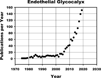
Since the 1940’s, it has been recognized that the endothelial surface layer (ESL) is comprised of a layer of polysaccharides, the glycocalyx, coated with adsorbed proteins that, together, comprise a barrier to blood cell contact with the endothelium and transvascular exchange of macromolecules. As noted in the figure below, interest in the endothelial glycocalyx grew steadily in the 70’s through 90’s with studies aimed to elucidate glycocalyx structure and function. However, with recognition in the early 2000’s that the ESL reflected a balance of the synthesis of new glycans and their shear dependent removal by activation of endothelial derived and circulating proteases and lyases, that interest rose exponentially. The importance of the glycocalyx as a mechanosensor for fluid shear stress was also recognized in the early 2000’s. More recently, the realization that the glycocalyx is degraded in vascular diseases (atherosclerosis, stroke, hypertension, diabetes, etc.) has led to a search for pharmaceuticals that can protect or regenerate the glycocalyx.
In view of the importance of the ESL in maintenance of hemostasis in health and disease, two symposia were held to review the state-of-the-art in knowledge of the structure of the glycocalyx, its role in the mechanotransduction of shear dependent changes in endothelial cell function, and its derangement in pathological disturbances such as diabetes and inflammation. To elucidate structural features that may play a role in mechanotransduction and transvascular exchange, Fan et al. have employed stochastic optical reconstruction microscopy (STORM) to examine the distribution of the principal glycosaminglycans (GAGs) heparan sulfate (HS) and hyaluronan (HA) on the EC surface. It is suggested that HS plays the major role in mechanosensing and that HA comprises the principal barrier to sieving of macromolecules. This paper is followed by the molecular modelling of Jiang et al. that aims to elucidate the relative movements of the core proteins and their associated sugar chains under fluid shear stresses. These studies reveal that fluid shear parallel to the EC surface results in three-dimensional movement of core proteins and they propose an alternate means of force transmission via the associated sugar chains. In the paper by Butler and Bhatnagar, it is proposed that the abluminal glycocalyx EC may participate in the mechanotransduction process by transmission of forces to focal adhesion complexes between the EC and its surroundings.
Analysis of the barrier function of the glycocalyx by Curry and Michel aims to elucidate the relationship between structure of the glycocalyx and its role as a barrier to transvascular exchange of macromolecules. Drawing upon observations of the exclusion of red cells and ultrafiltration of albumin, a model is developed that suggests that the ESL consists of a porous outer layer and a more selective membrane-associated inner layer where the albumin permeability coefficient and hydraulic conductivity in the outer layer are an order of magnitude larger than those of the inner layer. This paper is followed by an analysis by Harding et al. of the effects of disruption of glycocalyx barrier function in regions of glycocalyx damage. Examination of the glycocalyx structure on cultured ECs in a flow chamber under disturbed flow compared to that in uniform flow suggests that GAGs are significantly reduced in a disturbed flow (analogous to that observed in atherosclerotic regions in vivo) and adhesion of circulating cancer cells increased significantly. They conclude that therapeutic strategies to maintain glycocalyx structure in such situations may mitigate the disease processes of atherosclerosis and cancer cell metastasis.
To further elucidate the role of proteoglycans in the progression of the disease process, Moran et al. examine the relative roles of the HS proteoglycan core proteins, glypican-1 and syndecan-1, and the HA receptor CD44, on the surface of cancer cells as potential targets for pharmaceutical intervention to mitigate metastasis. They conclude that glypican-1 provides the mechanical linkage from HS to sense flow and initiate mechanotransduction leading to metastasis.
In the paper by Desideri et al. the relationship between degradation of the glomerular glycocalyx and onset of diabetic nephropathy is examined. Evidence is presented that treatment with paracrine growth factors can modify VEGFA signaling, rescue albumin permeability and restore the glomerular glycocalyx in models of diabetes. It is suggested that manipulation of VEGF receptor 2 signaling may protect and restore the glomerular glycocalyx in diabetic nephropathy. In a review article by Harris et al., it is suggested that blood flow distribution in the diabetic retina may be adversely affected by loss of the endothelial surface layer. Further, observations of elevated plasma heparanase, hyaluronidase, and hyaluronic acid, and increased concentrations of MMPs in the vitreous and plasma suggest a strong correlation between glycocalyx damage and retinopathy.
In the process of inflammation, the significance of shedding of proteoglycans and GAGs from the EC surface in inflammation is reviewed by Lipowsky. The loss of the glycocalyx in an animal model of inflammation is examined in light of enhancement of leukocyte-endothelium (WBC-EC) adhesion due to exposure of WBC adhesion receptors on the EC surface. New data is presented that reveals that activation of the EC to initiate glycan shedding results in a relatively rapid loss of HA that is followed later by shedding of HS. Based upon the structural observations of Fan et al. in this symposium, it is suggested that HA may be the principal barrier to WBC-EC adhesion, and that the HA layer near the EC surface is relatively stiffer compared to the HS layer on the glycocalyx surface.
Thus, the papers presented in this thematic issue of Biorheology cover a broad spectrum of functions of the EC glycocalyx that revolve around its structural and rheological properties.
Guest Editors:
John Tarbell, The City College of New York, USA and Hans Vink, Maastricht University, The Netherlands
Co-Editors-in-Chief
Herbert H. Lipowsky and Brian M. Cooke


